Functional capacity - knee pathology
The patient is a 21 year old male wrestler.
During a wrestling bout, while pinning his opponent with a "scissors" hold, he
claimed to have, "heard a popping noise and felt a pain in his right knee." The
patient complained of medial/lateral joint pain. The condition did not improve. Over the
course of 2 years, this patient was examined by 6 physicians [3 Orthopedic Surgeons].
Objective testing, including x-ray, MRI, and physical examination, failed to provide
evidence of true pathology; therefore, no further treatment was implemented. The last
Orthopedic Surgeon to examine the patient ordered a Functional Gait and Motion Analysis
Study. [see Illustration I]
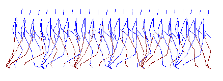
Illustration I
Side View - Gait Kinematics
Test Protocol
A test was designed to analyze the kinematics, kinetics and
functional electromyography of the patient's bilateral hips, knees and ankles. Video
computerized motion analysis, walking track force platform system and multi-channel
dynamic electromyography procedures were implemented to gather the necessary data.
Functional Motion Analysis Results
Kinematic analysis revealed an abnormality in the
distribution of vertical force loading, right knee, during the weight-bearing phase [see
Graph I]. This graph shows a distinct vertical pressure abnormality pattern for the right
knee. Note the mid-stance drop and increase of pressure. Further analysis shows the
loading abnormality to be specifically centered in the medial-posterior aspect of the
right knee [see Graph II].
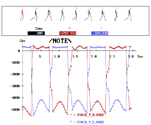
Graph I
Vertical Loading - Right vs. Left Knee
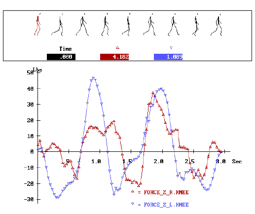
Graph II
Vertical Loading - Right vs. Left Knee
This result was further confirmed with kinetic force
platform analysis. EMG and dynamometer strength testing confirmed weakness in the right
hamstring muscle group.
Outcome
The functional motion analysis test results gave the
surgeon objective evidence and justification to proceed with Arthroscopic surgery. During
surgery of the right knee, the surgeon discovered a small tear in the region of the
posterior horn of the medial meniscus. The free fragments were removed. Post Arthroscopic
surgery, the patient was referred to physical therapy for 4 weeks. The patient reached his
strength and functional plateau and is back to unlimited functional activities, including
sports.