|
Marker Sets


Categories


|
 Contact
us Contact
us |
|
General info
+1 949 858 4216 |
|
Sales & support
+1 619 992 3089 |
|
E-mail
Information
Sales
Support |
|
CHECK OUT
[FrontPage Include Component]
|
| |
2.4 Marker sets
Check here for
currently supported marker sets by APAS/Gait version 1.04 - Revision 2004.10.21
.
 |
2D (meta, ankle, knee,
hip) (how to define the CF-files). |
|
KO (one or two legs) |
|
KM (one or two legs) |
|
HH (one or two legs) |
|
Anthropometric measures
and table values for 2D, KO, KM and HH |
|
Templates for missing
markers with 4, 5 and 6 cameras. |
For
gait kinematic and kinetic analysis, a number of markers
are
attached on
specific
locations of various body parts.
Markers are tracked automatically by optoelectronic system to be represented as
points in 3D space. After automatic tracking and 3D conversion, each marker has
its own positional information/data in GCS. The configuration of specific
locations of markers is called marker set. There are several conditions to be a
good marker set.
|
Easy
to track automatically
- Should minimize the chance of hiding or merging of the markers |
|
At
least three noncolinear markers on a body segment
- At least three markers are attached on a body segment
- Can be reduced to 2 markers if we use virtual markers, such as joint
center
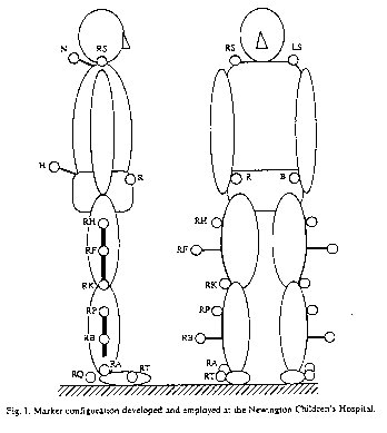
- for example, Helen Hayes marker set uses 13 or 15 markers for 7 body
segments. |
|
Able
to define anatomically relevant LCS
- To estimate joint centers accurately and to define anatomical planes
(sagittal or coronal) of body segments should be warranted. |
APAS/Gait can use
five marker sets. Four of those are the most widely
used. One is newly developed specifically for APAS/Gait.
Here
are a brief description and a comparison table of the five marker sets..
1. Original Helen Hayes(HHo) marker set that used by Davis and Kadaba.
2. Modified Helen Hayes(HHm) marker set
3. Original Kit Vaughan's marker set(KVo) - published on his 1st edition of
"Dynamic of human gait"
4. Modified Kit Vaughan's set (KVm) - published on his 2nd edition (CD ROM version)
5. Sun's marker set
* Comparison Table
* Marker sharing
** Marker Name and Position
3.2.5 Anthropometry
for marker sets (Anthro
for kinetics is not included)
* Comparison Table
|
HHo |
HHm |
KVo |
KVm |
Sun |
No. of markers |
15 (17 for static) |
15 (with two heels) |
15 |
15 |
19 (23 for static) |
Use of wand |
o |
o |
x |
o |
x |
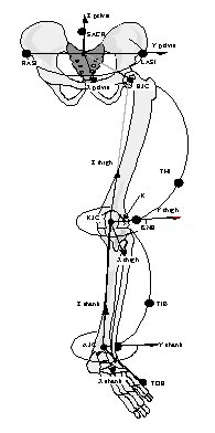
Hip joint center estim. |
Davis
algorithm |
Davis algorithm |
based on stero
x rays |
based on stero x rays |
Bell's algorithm |
Knee joint center estim. |
from thigh wand(marker) and knee width |
from thigh wand and knee width |
based on stero x rays |
from tibia wand and knee width |
midpoint btwn med. and lat.
epicondyle |
Ankle joint center estim. |
from tibia wand and knee width |
from tibia wand and knee width |
based on stero x rays |
based on stero x rays |
midpoint btwn med. and lat.
malleolus |
Potential problems
|
1. No heel markers-cannot
calculate Euler angles
(but for Dalmatian projec, heel markers
are used for HHo)
2. Difficulty to digitize G. trochant
marker
3. Swaying wand markers esp. during jerky gait
4. Possible inaccurate hip joint center estim |
1. Swaying wand markers
esp. during jerky gait
2. Possible inaccurate hip joint
center estim
3. Because there is no G troch. marker, "2"
can introduce error on knee joint center estim.
|
1. Somewhat difficult
to digitize automatically
2. Possible inaccurate hip, knee
and ankle joint center estim, because of small sized sample for
the stereo x rays. |
1. The hip
joint center estim. is same as KVo.
2. Use tibia markers to
estimate knee joint center.
3. Swaying wand markers esp.
during jerky gait |
1. Soft
tissue movement beneath the thigh ant. marker |
* No algorithm of hip joint center estimation seems to be absolutely accurate.
- more about hip joint center estimation
** Marker sharing
Marker
Name |
HHo |
HHm |
KVo |
KVm |
Sun |
SACR |
O |
O |
O |
O |
O |
R,L ASIS |
O |
O |
O |
O |
O |
R,L GTRO |
O |
- |
O |
- |
- |
R,L THI_W |
O |
O |
- |
O |
- |
R,L THI_L |
- |
- |
- |
- |
O |
R,L THI_A |
- |
- |
- |
- |
O |
R,L LCON |
O |
O |
O |
O |
O |
R,L MCON |
- |
- |
- |
- |
D
(for static) |
R,L TIB_W |
O |
O |
- |
O |
- |
R,L TTUB |
- |
- |
O |
- |
O |
R,L FH |
- |
- |
- |
- |
O |
R,L LMAL |
O |
O |
O |
O |
O |
R,L MMAL |
- |
- |
- |
- |
D
(for static) |
R,L MT |
D
(5th
MT) |
O |
D
(5th
MT) |
O |
O |
R,L HEEL |
O (-)* |
O |
O |
O |
O |
Totoal
No |
15 |
15 |
15 |
15 |
19 (21 static) |
*: In original
HHo, there
was no heel marker. But for this study we included heel markers for HHo marker
set. We are not to compare the ankle angle with or without heel markers because
it is obvious that former is better than later.
** Marker Name and Position
Marker
Name |
Full
Name |
Detailed
Instructions |
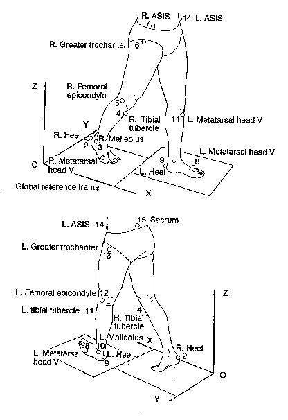
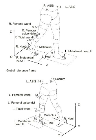
SACR |
Sacrum |
keep
the center of marker to be on the reversed direction of �i� vector
of pelvis LCS |
R,L ASIS |
Ant.
Sup. Iliac spine |
keep
the center of marker to be on the direction of �i� vector of pelvis
LCS |
R,L GTRO |
Greater
Trochanter |
On the
greater trochanter of femur |
R,L THI_W |
Thigh
wand |
| a stick marker on the
lower lateral thigh.
| Should be on the plane
that is formed by hip join, knee joint and lateral epicondyle
marker.
| |
|
R,L THI_L |
Thigh
lateral |
| on the lateral thigh.
| The plane formed by
RTHI_A, hip joint center and knee joint center will be sagittal
plane of thigh.
| |
|
R,L THI_A |
Thigh
anterior |
| on the anterior thigh.
| Should be on the plane
that is formed by hip join, knee joint and lateral epicondyle
marker.
| |
|
R,L LCON |
Lateral
Epicondyle |
| on the center of lateral
epicondyle of femur.
| You can find it more
easily if the subject�s knee flexed a little.
| |
|
R,L MCON |
Medial
Epicondyle |
| only for static trial.
| on the center of medial
epicondyle of femur.
| You can find it more
easily if the subject�s knee flexed a little.
| | |
|
R,L TIB_W |
Tibial
wand |
| a stick marker on the
upper lateral surface of lower leg.
| Around upper 1/3 point
of fibular head to lateral malleolus, where is no tibial torsion
component.
| |
|
R,L TTUB |
Tibial
tuberosity |
On the
tibial tuberosity |
R,L FH |
Fibular
Head |
| On the fibular head.
| But 2 or 3cm inferior
to fibular head would be better to avoid merging with lateral
condyle marker
| |
|
R,L LMAL |
Lateral
Malleolus |
Center
of lateral malleolus |
R,L MMAL |
Medial
Malleolus |
Center
of medial malleolus |
R,L MT |
Head
of 2nd Metatarsal |
on the
second MT head |
R,L HEEL |
Heel |
| On heel.
| The line between RHEEL
and RMT should be parallel to the ground. To achieve this condition:
one should measure the thickness(height) of 2nd MT
head(=RMTHth). The RHEEL should be above from
the ground with the amount of m_offset1+RMTHth.
| |
|
3.2.5 Anthropometry
for marker sets (Anthro
for kinetics is not included)
|
