THE UNIVERSITY OF CALGARY
Three-Dimensional Tibiocalcaneal and Tibiofemoral Kinematics During Human Locomotion - Measured with External and Bone Markers
by
Christoph Reinschmidt
A DISSERTATION
SUBMITTED TO THE FACULTY OF GRADUATE STUDIES
IN PARTIAL FULFILLMENT OF THE REQUIREMENTS FOR THE
DEGREE OF DOCTOR OF PHILOSOPHY
DEPARTMENT OF MEDICAL SCIENCE
CALGARY, ALBERTA
MARCH, 1996
Christoph Reinschmidt 1996
FACULTY OF GRADUATE STUDIES
The undersigned certify that they have read, and recommend to the Faculty of Graduate Studies for acceptance, a dissertation entitled "Three-Dimensional Tibiocalcaneal and Tibiofemoral Kinematics During Human Locomotion - Measured with External and Bone Markers" submitted by Christoph Reinschmidt in partial fulfillment of the requirements for the degree of Doctor of Philosophy.
Supervisor, Dr. B.M. Nigg
Department of Medical Science
Dr. A.J. van den Bogert
Faculty of Kinesiology
Dr. C.B. Frank
Department of Surgery
Dr. J.L. Ronsky
Department of Mechanical Engineering
External Examiner, Dr. P.R. Cavanagh
The Center of Locomotion Studies
Pennsylvania State University, U.S.A.
Date
Three-dimensional kinematics of the lower extremities are typically assessed with external markers attached to the segments of interest. However, these markers may move considerably with respect to the underlying bone, and thus, large errors may be introduced. The purpose of this study was to determine the three-dimensional skeletal tibiocalcaneal (ankle joint complex, AJC) and tibiofemoral (knee) motion during the stance phase of walking and running, and to compare this to the respective motion determined from external markers.
Marker triads were attached to intracortical bone pins inserted into the calcaneus, tibia, and femur of five subjects. Additionally, external markers were attached to the shoe, shank, and thigh. For each subject, three walking and five running trials were filmed with three high speed cameras (50 Hz for walking, 200 Hz for running). Cardan angles were calculated to express the intersegmental knee and AJC motion. Knee flexion/extension, ab/adduction, and internal/external knee rotation as well as AJC plantar/dorsiflexion, ab/adduction and in/eversion were calculated both from skeletal and external markers.
For walking and running, it was found that the skeletal tibiocalcaneal (AJC) motions were well reflected when using external markers. However, the rotations were generally overestimated when using external markers. For instance, during running, the maximal initial eversion occurring from touchdown to midstance averaged 16.0 when using external markers. However, the same variable determined from skeletal markers was only 8.6.
During walking and running, the skeletal knee flexion/extension was well represented with skin markers. For ab/adduction and internal/external knee rotation, the agreement between external and skeletal kinematics ranged from good to virtually no agreement. In some subjects, the errors exceeded the actual skeletal motion. Methodological problems were also identified with the determination of tibiofemoral kinematics. Internal/external knee rotation and particularly ab/adduction can be expected to be small, and thus, they are highly affected by cross-talk caused by uncertainties in defining the anatomical coordinate systems.
The results of this project suggest that (a) tibiocalcaneal motions are generally well represented with external markers, but absolute values have to be interpreted with caution, and that (b) knee rotations other than flexion/extension may be affected with substantial errors when using skin markers.
Chapters 3, 4, and 5 of this thesis are based on the following manuscripts:
- Reinschmidt, C., Bogert, A.J. van den, Lundberg, A., Murphy, N., Nigg, B.M., Stacoff, A., and Stano, A. Tibiofemoral and tibiocalcaneal motion during walking: skin vs. bone markers. Submitted to Gait & Posture.
- Reinschmidt, C., Bogert, A.J. van den, Murphy, N., Lundberg, A., and Nigg, B.M. Tibiocalcaneal motion during running - measured with external and bone markers. Submitted to Clin. Biomech.
- Reinschmidt, C., Bogert, A.J. van den, Nigg, B.M., Lundberg, A., and Murphy, N. Effect of skin movement artefact on the calculation of knee joint motion during running. Submitted to J. Biomech.
This thesis has been written as a compilation of (stand-alone) papers arranged in chapters. Since the rational and methods of these papers are similar, some chapters (chapters 3 to 5) contain redundant information, particularly, in the "introduction" and "methods" sections. Additionally, portions of the general introduction and literature review of the thesis can be found in the introduction of chapters 3 to 5.
I would like to express my sincere gratitude and appreciation to the following individuals and institutions. Without their help this thesis would not have been possible.
- Dr. Benno M. Nigg, for his help, guidance, encouragement and support throughout my Ph.D. marathon.
- Dr. Ton van den Bogert, who was my "personal navigator" in the three-dimensional space, and who always had time to discuss the technical aspects of my thesis.
- Drs. Cy Frank, Peter Cavanagh, and Janet Ronsky for serving on my thesis committee.
- Dr. Arne Lundberg, who with his cool Swedish manner handled "with ease" all the medical aspects of the experiments.
- Andrzej (Super-) Stano, Dr. Norman Murphy, Alex Stacoff, and Anna Josephson for their help during the experiments in Huddinge, Sweden.
- Drs. Stig Drevemo and Christopher Johnston of the Swedish University of Agricultural Science in Uppsala for providing part of their movement analyses equipment.
- Alex Stacoff and Dr. Edgar Stssi, who introduced me into the field of biomechanics when I first started working as a research assistant at the Biomechanics Laboratory at the ETH Zrich.
- All fellow graduate students and current as well as former members of the Human Performance Laboratory for great talks, discussions, social hours, and great Volleyball games.
- Evelyne for "luring" me to Calgary.
- Sandro Nigg, the professional digitizer, for accurately and meticulously digitizing most of the films.
- All the subjects who participated in the invasive experiments of my thesis.
- The following institutions for their financial support: the Swiss Federal Sports Commission (ESK), the ADIDAS sport shoe company, the Kanton Aargau of Switzerland (Zentralstelle fr Ausbildungsfrderung des Kantons Aargau), the Olympic Oval Endowment Fund of Calgary, the Swedish Defence Materials Administration, and the Going Global 1992 Fund of Canada.
to my parents, Elsa and Gusti, and to Evelyne
Approval page
*ABSTRACT
PREFACE
ACKNOWLEDGEMENTS
DEDICATION
TABLE OF CONTENTS
LIST OF TABLES
LIST OF FIGURES
CHAPTER 1: Introduction
Purpose
Relevance
CHAPTER 2: Review of Literature
Ankle-Joint Complex and Knee Joint Motion
Ankle-joint complex motion
Walking
Running
Knee (tibiofemoral) joint motion
Walking
Running
Determination of three-dimensional intersegmental motion
Direct measurements of skeletal motion
Bone pin studies
External fixator devices
Percutaneous skeletal tracker
Roentgen-stereo analysis
Video fluoroscopy
Determination of skin movement artefact
Methods to reduce the skin movement artefact
Skin frames
Solidification model
Marker arrays, clusters
Correction algorithm
Shoe windows
CHAPTER 3: Tibiofemoral and Tibiocalcaneal Motion During Walking: Skin vs. Bone Markers
Introduction
Methods
Subjects
Surgical procedure
Marker placements
Experimental protocol and set-up
Motion recordings
Three-dimensional reconstruction
Coordinate Transformations
Reference frames and relative orientation
Segmental error contributions
Assumptions and limitations
Results
Effect of the pins
Accuracy of spatial reconstruction
Tibiofemoral Motion
Variability
Knee ab/adduction
Internal/external rotation
Flexion/extension
AJC Motion
Variability
In/eversion
Ab/adduction
Plantar/dorsiflexion
Discussion
Tibiofemoral Motion
AJC Motion
Conclusions
Summary
CHAPTER 4: Tibiocalcaneal Motion During Running - Measured With External And Bone Markers
Introduction
Methods
Bone and skin markers
Protocol and motion recordings
Data analysis
Variables
Results
Effect of pins on running kinematics
Tibiocalcaneal rotations
External vs. bone marker based rotations
Maximal eversion
Discussion
Tibiocalcaneal rotations
External vs. bone marker based rotations
Segmental error analysis
Maximal eversion
Conclusions
Summary
CHAPTER 5: Effect of Skin Movement Artefact on the Analysis of Knee Joint Motion During Running
Introduction
Methods
Results
Effect of bone pins
Accuracy of spatial reconstruction
External vs. skeletal kinematics
Segmental error
Discussion
Summary
CHAPTER 6: Methodological Considerations and "Normal" Tibiofemoral Joint Motion in Running
Stability of Femur Pin Attachment
Introduction
Methods
Results and Discussion
Anatomical Coordinate Systems and Crosstalk
Cross-talk
Tibiofemoral Joint Motion During Running
Methods
Results and Discussion
Rotations
Translations
Summary and Conclusions
CHAPTER 7: Implications for Future Studies Using External Markers
Shoe/Foot (Calcaneus)
Shank (Tibia)
Methods
Results and Discussion
Thigh (Femur)
Quantitative Analysis of Movement Artefact of Thigh Markers
Methods
Results and Discussion
Qualitative Analysis of Movement Artefact of Thigh Markers
Recommendations
Conclusions and Future Research
CHAPTER 8: Summary and Conclusions
REFERENCES
APPENDIX: Methods for the Calculation of Rigid Body Kinematics
NOTATIONS
ANATOMICAL REFERENCE FRAMES
Definition of anatomical coordinate system based on a neutral position
Definition of anatomical coordinate system based on RSA
FILM/ VIDEO ANALYSIS
Absolute orientation of a segment
Relative orientation of two adjacent segments
CALCULATION OF CARDAN ANGLES AND TRANSLATIONS
Tibio-femoral motion
Tibiocalcaneal motion
Rotation Matrices
LIST OF TABLES
Table 1: Findings of studies investigating the discrepancy between external and skeletal marker based kinematics.
Table 2: Mean residuals of the DLT calculations for the femur markers and the thigh markers (Th2 to Th6) averaged over the stance phase of the three walking trials.
Table 3: Root mean square (RMS Diff.) and maximal difference (Max. Diff.) between skin and bone marker based knee rotations during the stance phase of walking, and qualitative agreement between the shape of the knee rotation curves derived from skin and bone markers. Note that all values were calculated from the average curves of the corresponding subjects.
Table 4: Root mean square (RMS Diff.) and maximal difference (Max. Diff.) between skin and bone marker based AJC rotations during the stance phase of walking, and qualitative agreement between the shape of the AJC rotation curves derived from skin and bone markers. Note that all values were calculated from the average curves of the corresponding subjects.
Table 5: Root mean square (RMS Diff.) and maximal difference (Max. Diff.) between skin and bone marker based tibiocalcaneal rotations during the stance phase of running expressed in absolute () terms, as well as relative (%) to the range of motion of the corresponding rotation. Values were calculated from the average curves of the corresponding subjects.
Table 6: Maximal change in tibiocalcaneal eversion from touchdown to maximal eversion during the stance phase of running calculated based on bone (
Table 7: Mean residuals of the DLT calculations for the femur (F1 to F3) thigh markers (Th2 to Th6) for the subjects averaged over the stance phase of the five running trials.
Table 8: Qualitative agreement between the shape of the skeletal and skin marker based knee rotations during the stance phase of running, and root mean square (RMS Diff.) and maximal difference (Max. Diff.) between skin and bone marker based knee rotations during stance, expressed in absolute () terms, as well as relative (%) to the range of motion of the corresponding rotation. Values were calculated from the average curves of the corresponding subjects.
Table 9: Root mean square (RMS Diff.) and maximal difference (Max. Diff.) between skin and bone marker based rotations of the tibia about its longitudinal axis (with respect to the laboratory coordinate system) during the stance phase of running expressed in absolute () terms, as well as relative (%) to the range of motion. Values were calculated from the average curves of the corresponding subjects (Fig. 24).
Table 10: Average error contribution of the thigh (mean thigh error, MTE) during running for different thigh marker combinations and for a skin frame. The placement of the thigh markers are depicted in Fig. 1.
Table 11: Average relative marker movement (RMM) of each thigh marker with respect to a femur (bone marker) fixed coordinate system. The RMM values were averaged from five running trials of the three subject for which valid femur pin data was available. The placement of the thigh markers are depicted in Fig. 1. * Values are likely to be affected by inaccuracies in the spatial reconstruction (DLT).
LIST OF FIGURES
Fig. 1: Bone (femur: F1 to F3; tibia: T1 to T3; calcaneus: C1 to C3), skin (thigh: Th1 to Th6; shank: S1 to S6) and shoe marker (Sh1 to Sh3) placements.
Fig. 2: Experimental set-up
Fig. 3: Effect of bone pins on skin marker based knee rotations during walking for one subject. Solid lines ( ) represent trials without bone pins, dashed lines (---) represent trials with pins. The averages of the three trials are displayed with thick lines. Movements labeled on the y-axis indicate rotational movements in the positive direction of the y-axis.
Fig. 4: Norm of residuals of the spatial reconstruction (direct linear transformation) displayed for all the markers for the 3 subjects with valid femur pin data. For each marker the mean ( SD) residuals of the 3 walking trials during the stance phase are plotted. The residuals are given in units of the digitizing board. 10 units correspond to approximately 3 mm. The order in which the markers are displayed for a given segment (e.g. thigh) corresponds to the order displayed in Fig. 1 (e.g. Th1 to Th6).
Fig. 5: Knee joint rotations during walking based on bone (femur, tibia) markers and external (thigh, shank) markers. Solid lines ( ) represent bone pin based kinematics, dashed lines (---) represent skin marker based kinematics. The averages of the three trials are displayed with thick lines. Movements labeled on the y-axis indicate rotational movements in the positive direction of the y-axis. Vertical lines indicate times where only 2 cameras were available.
Fig. 6: Ankle joint complex rotations during walking based on bone (tibia, calcaneus) markers and external (shank, shoe) markers. Solid lines ( ) represent bone pin based kinematics, dashed lines (---) represent skin (shoe) marker based kinematics. The averages of the three trials are displayed with thick lines. Movements labeled on the y-axis indicate rotational movements in the positive direction of the y-axis. Vertical lines indicate times where only two cameras were available.
Fig. 7: Effect of the skin movement artefact of the thigh ( ) and shank (---) on knee rotations during the stance phase of walking. Each curve represents the average difference between the bone based knee motion (femur-tibia) and the thigh-tibia ( ) as well as the femur-shank (---) motion in one subject. Positive error values indicate overestimation of the bone movements due to the skin movement artefact.
Fig. 8: Effect of the skin movement artefact of the shoe ( ) and shank (---) on rotations at the ankle joint complex during the stance phase of walking. Each curve represents the average difference in one subject between bone based AJC motion (femur-tibia) and the shoe-tibia ( ) as well as the calcaneus-shank (---) motion. Positive error values indicate overestimation of the bone movements due to the skin movement artefact.
Fig. 9: Effect of bone pins on surface marker based tibiocalcaneal rotations during the stance phase of running for two subjects. Solid lines ( ) represent the three trials without bone pins (pre-operative trials), dashed lines (---) represent the five trials recorded with pins. The averages of the single trials are displayed with thick lines. Movements labeled on the vertical axis indicate rotational movements in the positive direction of the vertical axis.
Fig. 10: Tibiocalcaneal rotations during the stance phase of running based on bone (tibia, calcaneus) markers and external (shank, shoe) markers. Solid lines ( ) represent bone pin based kinematics, dashed lines (---) represent skin/shoe marker based kinematics. The averages of the five trials are displayed with thick lines. Movements labeled on the vertical axis indicate rotational movements in the positive direction of the vertical axis.
Fig. 11: Maximal eversion (during the stance phase of running) calculated from skeletal motion (
Fig. 12: Errors in tibiocalcaneal rotations (during the stance phase of running) due to relative movement between external markers on the shoe ( ) with respect to the calcaneus, and between external markers on the shank (---) with respect to the tibia. Each curve represents the average subject difference between bone based tibiocalcaneal motion and the tibia-shoe ( ) as well as the shank-calcaneus (---) motion. Positive error values indicate overestimation of the bone movements due to the skin and shoe movement artefact.
Fig. 13: Effect of bone pins on external marker based tibiofemoral rotations during the stance phase of running. Solid lines ( ) represent the three trials without bone pins (preoperative trials), dashed lines (---) represent the five trials recorded with pins. The averages of the single trials are displayed with thick lines. Labels on the vertical axis indicate knee movements in the positive direction of the vertical axis.
Fig. 14: Norm of residuals of the spatial reconstruction (DLT) displayed for all markers for the 3 subjects with valid femur pin data. For each marker the mean ( SD) residuals of the 5 running trials during the stance phase are plotted. The residuals are in units of the digitizing board. 10 units correspond to approximately 3 mm. The order in which the markers are displayed for a given segment (e.g. thigh) corresponds to the order displayed in Fig. 1. Note that for completeness the residuals of the shoe and calcaneus markers were also included in this figure even though these markers were not used for the results presented in this chapter (but for the results in chapter 4).
Fig. 15: Tibiofemoral rotations based on bone (tibia, femur) markers and skin (shank, thigh) markers during the stance phase of running. Solid lines ( ) represent bone pin based kinematics, dashed lines (---) represent skin marker based kinematics. The averages of the five trials are displayed with thick lines. Movements labeled on the vertical axis indicate rotational movements in the positive direction of the vertical axis.
Fig. 16: Effect of the skin movement artefact at the thigh ( ) and at the shank(---) on tibiofemoral rotations during the stance phase of running. Each curve represents the average subject difference between the skeletal tibiofemoral motion and the tibia-thigh ( ) as well as the shank-femur (---) motion. Positive error values indicate overestimations of the bone movements due to the skin movement artefact.
Fig. 17: Protocol for the motion measurements for each subject.
Fig. 18: Knee (tibiofemoral) positions during various standing trials displayed in chronological order for subject 1, 3, and 5. Within subjects, the alignment was always calculated with respect to standing trial 2 which was used to define the neutral knee positions for all running trials (chapter 5), and which was recorded immediately prior to these running trials. Standing trial 1 is the standing trial used for the walking trials (chapter 3). Standing trials 3 to 8 are standing trials which were part of another study not reported in this project. (Standing trial 5 belongs to barefoot running trials; standing trials 3, 4, 6, 7, and 8 to running trials for different shoe conditions).
Fig. 19: Femur positions during various standing trials displayed in chronological order for subject 1, 3, and 5. Within subjects, the alignment was always calculated with respect to the femur position recorded for the standing trial 2. For a description of the standing trials see Fig. 18.
Fig. 20: Tibia positions during various standing trials displayed in chronological order for subject 1, 3, and 5. Within subjects, the alignment was always calculated with respect to the tibia position recorded for the standing trial 2. For a description of the standing trials see Fig. 18.
Fig. 21: Points digitized in roentgen-stereographic pictures in order to establish anatomical coordinate systems for the tibia and femur. The points identified are similar to the ones digitized by Lafortune et al. (1992a).
Fig. 22: Skeletal tibiofemoral rotations during the stance phase of running. Solid lines ( ) represent joint kinematics calculated using standing trials to define the anatomical coordinate systems, dashed lines (---) represent kinematics based on coordinate systems determined from RSA. Averages of the five trials (thin lines) are displayed with thick lines. Labels on the vertical axes indicate rotational movements in the positive direction of the vertical axes. Note that the solid lines presented in this graph are the same as the solid lines displayed in Fig. 15.
Fig. 23: Skeletal tibiofemoral translations during the stance phase of running of one subject. Averages of the five single trials (thin lines) are displayed with thick lines. Labels on the vertical axes indicate joint translational movements in the positive direction of the vertical axes. Note that the change in movement should be considered rather than the absolute values displayed on the vertical axes, since also in the "neutral" position the origin of the tibial and femoral reference frames are already at same distance apart from each other.
Fig. 24: Rotation of the tibia about its longitudinal axis with respect to the laboratory coordinate system during the stance phase of running. Tibial rotation is displayed for the five subjects both based on skeletal ( ) and skin (---) markers. Thin lines indicate single trials, thick lines the average of the five single trials.
Fig. 25: Thigh frame thought to reduce the effect of the skin movement artefact with respect to the skeletal tibiofemoral rotations
Quantitative kinematic analysis of the lower extremities during human locomotion is an important tool for a thorough understanding of normal and pathological functions of the joints of the lower extremities. Until a few decades ago, kinematic assessments of human joints were confined to two-dimensional or pseudo three-dimensional (e.g. Levens et al., 1948) analyses. However, during the last two to three decades, three-dimensional analyses have become common with advances in photogrammetric techniques and with increased computer power and knowledge enabling the collection of spatial data in an automatic or semi-automatic manner. However, accurate determination and advances in the kinematic assessment of joints and joint-complexes have been hindered by the fact that surface markers may not give an accurate representation of the kinematics of the underlying bone during locomotion.
In routine kinematic analyses of the lower extremities (foot/shoe, lower and upper leg), skin markers or skeletal linkages attached to the segments of interest are typically used to represent the movement of the underlying bone. However, large errors may be introduced as a result of the relative movement between skin and underlying bone. This is of particular concern if the kinematics are assessed during a highly dynamic movement such as running, and if the segments of interest consist of a substantial amount of soft tissue such as the thigh. This source of error, typically referred to as the skin movement artefact, is believed to be the most important error in current human movement analyses (Cappozzo et al., 1996).
One way to avoid the problem inherent with surface markers is to directly measure skeletal motion of the respective segments. Different methods have been used to directly measure in vivo skeletal motion. They include stereo radiography (Lundberg, 1989; Maslen and Ackland, 1994), bone pins (Levens et al., 1948; Karlsson, 1990; McClay, 1990; Murphy, 1990; Lafortune et al., 1992a; Lafortune et al., 1994), external fixation devices (Cappozzo et al., 1996; Andriacchi and Toney, 1995), and a percutaneous skeletal tracker (Holden et al., 1994a). However, the applicability of such methods is limited, mainly due to the invasiveness of such procedures. Consequently, routine kinematic gait analysis used for clinical assessment must rely on measurements based on superficial skin markers. Therefore, knowledge about the skin movement artefacts is crucial for the interpretation of kinematic results based on external markers, in particular, if the results are used to decide on strategic interventions such as surgical procedures or to assess the success of an intervention.
In recent years, several studies have been published investigating the skin movement artefact at the lower extremities. For slow or quasi-dynamic movements, a substantial amount of skin movement artefact was found (Lafortune et al., 1992b; Cappozzo et al., 1996; Karlsson and Lundberg, 1994; Maslen and Ackland, 1994). For instance, Karlsson and Lundberg (1994) showed that skin markers at the thigh moved as much as 4 cm with respect to the underlying femur during a longitudinal knee rotation. They reported misreadings of up to 20 in tibiofemoral joint rotations caused by the skin movement artefacts at the femur and tibia.
During gait, the skin movement artefacts have been investigated using various methods. Karlsson (1990) and Murphy (1990) compared skin and bone pin mounted marker arrays at the femur and tibia. They concluded that the skin mounted marker arrays did not accurately reflect the tibiofemoral motion, especially during the stance phase of walking. Karlsson (1990) and Murphy (1990) expressed tibiofemoral joint motion in terms of instantaneous helical axes, a concept not widely used in gait analysis. The results of their investigations are therefore of limited value for general gait analysis studies. Angeloni et al. (1992) and Cappozzo et al. (1996) used patients with external fracture fixation devices to determine the relative movement between skin markers and the fixation device. They reported that markers placed on bony landmarks moved 1 cm to 2 cm with respect to the underlying bone. However, they did not report the effect of this relative movement on the measurement of knee rotations, and additionally, the gait of these patients may not have been normal. Recently, Holden and co-workers (1994a) developed a "percutaneous skeletal tracker", a rigid device fixed to the tibia and fibula by means of small bone pins. They reported that the greatest differences in translation and rotation occurred along and around the longitudinal shank axis, and that the relative displacements between skin markers and underlying bone were reproducible within subjects, but they varied across subjects. Their study was confined to the shank where much less skin movement artefact can be expected compared to the thigh. Although several studies have been presented looking at the skin movement artefact during walking, to date no comprehensive studies have been published determining the effect of the skin movement artefact on the three-dimensional knee rotations during walking. Moreover, no results have ever been presented regarding the effect of skin movement artefact during a highly dynamic movement, such as running, where the effect of the skin movement artefact can be expected to be fairly high.
Besides reporting the skin movement artefact, several studies have also investigated the possibility of minimizing the skin movement artefact with different methods. Ronsky and Nigg (1991) and Angeloni and co-workers (1993) proposed the use of rigid skin frames in order to minimize the skin movement artefact. Markers attached to skin frames appeared to provide better results than markers directly attached to the skin (Ronsky and Nigg, 1991; Angeloni et al., 1993). Karlsson (1990) also showed that the type of mounting has an influence on the amount of skin movement artefact. Recently, Chze and co-workers (1995) suggested another approach to reduce the skin movement artefact. They proposed a solidification model for a set of segmental markers in order to reduce the skin movement artefact. Such a "best rigid model" appears to mainly decrease the motion of the markers relative to each other. However, the three (or more) markers providing the best rigid model (Chze et al., 1995) may still move as a whole with respect to the underlying bone. Therefore, a considerable amount of skin movement artefact may still be present after applying such a procedure. Another approach using a large (redundant) number of markers per segment has been suggested by Andriacchi and Toney (1995). They showed that the knee joint kinematics during a stepping-down movement could be accurately determined when using marker clusters (10 markers at each segment) and eigenvector based calculations. However, the high number of markers at each segment may be critical for automatic spatial tracking systems and it remains to be shown whether the proposed procedure would also be successful when determining the knee joint kinematics during highly dynamic movements such as running. Another approach to minimize skin movement artefacts consists of models reducing the relative motion of skin and underlying bone via the use of correction algorithms. Such a model has successfully been developed for two-dimensional equine gait analysis (Bogert et al., 1990; van Weeren et al., 1992). Before such a model can be developed for humans, more knowledge is needed about the relative skin movement at the foot/shoe, shank, and thigh.
Although there have been a number of studies investigating the skin movement artefact during gait in recent years, no comprehensive studies have been published that looked at the effect of the skin movement artefact on both the three-dimensional knee (tibiofemoral) joint kinematics and the three-dimensional ankle joint complex, AJC, (tibiocalcaneal) kinematics during human locomotion, i.e. during both walking and running.
Therefore, the purposes of this thesis are:
- to determine the three-dimensional skeletal tibiocalcaneal motion during the stance phase of walking and to compare it to tibiocalcaneal motion determined from external markers,
- to determine the three-dimensional skeletal tibiofemoral motion during the stance phase of walking and to compare it to tibiofemoral motion determined from external markers,
- to determine the three-dimensional skeletal tibiocalcaneal motion during the stance phase of running and to compare it to tibiocalcaneal motion determined from external markers,
- to determine the three-dimensional skeletal tibiofemoral motion during the stance phase of running and to compare it to tibiofemoral motion determined from external markers.
These specific purposes will be addressed in the following chapters. Purposes a) and b) will be accomplished in Chapter 3 where the methods of this project will be described in detail. Purpose c) will be addressed in Chapter 4, and purpose d) in Chapter 5. Additionally, methodological considerations and problems associated with this project will be presented in Chapter 6.
Kinematic analyses of the lower extremities using external markers rely on the assumption that bone kinematics are well represented with the use of these externally mounted markers. However, knowledge about the skin movement artefact during human locomotion is limited, and thus, it is not known whether or not it is fair to assume that the bone kinematics during human locomotion are well reflected when employing external markers. The results of this project will show how well externally measured kinematics actually agree with bone kinematics during human locomotion (walking and running). This information will help to (re)interpret the results of (previous) kinematic studies that employed external markers. Knowledge about the magnitude of the error in joint kinematics due to the skin movement artefact will also help in the design of future studies using external markers.
The results of this project will also provide information about the "normal" tibiocalcaneal and tibiofemoral motion during walking and running. To date, skeletal tibiocalcaneal kinematics have never been reported, either during walking or during running. Therefore, this project will provide unique data which may help to gain a better understanding of the functioning of the knee and ankle-joint complexes. This information may also be used as input for simulations of walking and running.
CHAPTER 2: Review of Literature
This review of literature is divided into five parts. In the first section, the current knowledge regarding ankle-joint complex (AJC) and knee joint motion during walking and running will be summarized. The second part deals with the methodology concerning the determination of three-dimensional intersegmental motion. In the third section, the literature and methods concerning direct measurements of skeletal motion will be discussed. In the fourth section, the current knowledge about the magnitude of the skin movement artefact will be summarized. The last part of this literature review will present and discuss methods that have been suggested to reduce the skin movement artefact.
Ankle-Joint Complex and Knee Joint Motion
This review of selected articles will focus on three-dimensional (angular) kinematic variables measured in-vivo during the stance phase of walking and running.
The human ankle-joint complex (AJC), linking the foot to the shank, has functionally been divided into two separate joints: the talocrural (or ankle) joint between shank (tibia and fibula) and talus, and the talo-calcaneo-navicular (or subtalar) joint (Inman, 1976). Due to the difficulty of measuring the movement of the talus with external markers, the entire ankle-joint complex has typically been simplified as a ball-and-socket joint with the Cardanic angles plantar/dorsiflexion, ab/adduction, and in/eversion (e.g. Soutas-Little et al., 1987; Areblad et al., 1990; Kepple et al., 1990; Moseley et al., 1996). The movement of the talocrural joint may be reasonably well represented by the plantar/dorsiflexion of the foot with respect to the leg since the axis of the talocrural joint is close to the medio-lateral axis (Inman, 1976) around which plantar/dorsiflexion is typically calculated. On the other hand, the average subtalar joint axis is inclined 42 (SD 9) from the horizontal plane, and deviated medially by 23 (SD 11) (Inman, 1976). Due to the obliqueness of this axis, the movement of the subtalar joint contains all three Cardanic angles. Hence, the rotation at the subtalar joint cannot be measured directly. In the past, the in/eversion measured either two or three-dimensionally has typically been used as an indicator of the rotation occurring around the subtalar joint axis (supination/pronation).
Plantar/Dorsiflexion. Probably the most comprehensive study on normal three-dimensional rearfoot kinematics has recently been published by Moseley and co-workers (1996). They determined the rearfoot kinematics of 14 subjects during barefoot walking using a stringent calibration system to standardize the neutral position across subjects. The average plantar/dorsiflexion curves showed that after heel contact, the rearfoot rapidly plantarflexed to a maximum (plantarflexion position) of 6.8 ( 1.3) occurring at 17% of stance phase. A progressive rearfoot dorsiflexion then took place until around 80% of stance, when a rapid plantarflexion occurred until take-off. These patterns of plantar/dorsiflexion agreed well with earlier studies (e.g. Kepple et al., 1990). However, the range of motion as well as the absolute positions at given percentages of stance may vary across studies determining the plantar/dorsiflexion of the foot with respect to the lower leg (Kadaba et al., 1990; Kepple et al., 1990; Moseley et al., 1996). For instance, the amount of initial plantarflexion motion extracted from average curves ranged between 3 (Kadaba et al., 1990) and 9 (Moseley et al., 1996). Such differences may be attributed to the different methods applied in these studies, such as different marker placements and differences in defining the anatomical foot and shank reference frames.
Ab/Adduction (int./external tibial rotation)*. Investigations regarding 3-D analysis of ab/adduction (or internal external tibial rotation) are limited. Kepple et al. (1990) reported large variability in ab/adduction patterns across the five subjects included in their study. In contrast, Moseley et al. (1996) found a consistent ab/adduction pattern across subjects. They found that after heel strike, a gradual increase in abduction took place, reaching a maximum abduction position (7) at 63% of stance phase. During the later stage of stance, an adduction movement took place and at toe-off the foot was in an adducted position of around 3 (Moseley et al., 1996).
In/Eversion. Controversial findings exist regarding the in/eversion patterns during the initial phase of stance. The results presented by Kepple et al. (1990) suggest that a sudden inversion occurs just after heel strike, while the results of other studies (Moseley et al., 1996; Delozier et al., 1991) suggest that a gradual eversion takes place from touchdown to midstance. However, apart from this inconsistency, the results of these studies appear to agree. The maximum eversion position is reached at around 60% of stance phase, when the foot starts to make an inversion motion until toe-off.
It is speculated that the initial sudden inversion reported by Kepple et al. (1990) was the result of "cross-talk" (see also chapter 6). The presented in/eversion pattern is very similar to the plantar/dorsiflexion pattern. Uncertainties in defining an anatomical coordinate system (e.g., for the definition of neutral, the subjects may have been in a slightly abducted position) may result in cross-talk in the sense that some of the plantar/dorsiflexion motion may be "picked up" by the in/eversion or ab/adduction. A similar phenomenon was encountered when comparing the initial eversion patterns between heel-toe and toe running (Reinschmidt et al., unpublished data). These running styles are characterized by distinct differences in the initial plantar/dorsiflexion movements. Similar differences were also present in the in/eversion of some subjects. It was speculated that the differences in in/eversion between the two running styles may have been partially caused by cross-talk due to inaccurate definitions of the anatomical foot and lower leg coordinate systems. However, further research is needed to substantiate these speculations.
Knowledge about the three-dimensional AJC motion is limited. No study including a large number of subjects has been conducted establishing "normal" AJC rotations for running.
Plantar/Dorsiflexion. The plantar/dorsiflexion motion at the ankle-joint complex during running stance is highly dependent on the type of striking pattern, i.e. rearfoot versus mid-foot and forefoot striking (McClay and Manal, 1995; Soutas-Little et al., 1987). Heel-toe runners, or in other words rearfoot strikers, exhibit an initial plantarflexion movement. After around 20% of stance, a dorsiflexion movement occurs which levels off at around 50% of stance. Towards the end of stance, a relatively fast plantarflexion movement takes place (Areblad et al., 1990; Soutas-Little et al., 1987). The patterns for midfoot strikers are similar, except for the initial 20% of stance. In contrast to rearfoot strikers, who exhibit an initial plantarflexion as the forefoot is lowered, midfoot strikers show a gradual dorsiflexion movement immediately following touch-down. Interestingly, it appears that in Soutas-Little et al. (1987), the exemplary curves for the runners exhibiting a heel and midfoot striking pattern were interchanged.
Ab/Adduction (int./external tibial rotation). To the author’s knowledge, ab/adduction and tibial (leg) rotation curves have only been reported by Soutas-Little et al. (1987) and Nigg et al. (1993), respectively. The two subjects, one rearfoot and one forefoot striker, in the study of Soutas-Little et al. (1987) showed similar patterns: A gradual abduction in the order of magnitude of 15 from touch-down to midstance was followed by an adduction movement in the order of magnitude of 10 during the second half of stance. The values for internal/external leg (tibial) rotation reported by Nigg and co-workers (1993) showed a larger range of motion than the exemplary curves for ab/adduction presented by Soutas-Little et al. (1987). An initial internal leg rotation took place from 0% to around 55% of stance averaging 21.8(n=30) (Nigg et al., 1993). During the second phase of stance, an external tibial rotation occurred averaging 19.
In/Eversion. In/eversion has gained by far the most attention in kinematic analyses of the ankle-joint complex. The main reason for this may be that certain in/eversion patterns, such as excessive eversion (pronation), have been linked to the incidence of a number of running injuries, such as Achilles tendinitis (Clement et al. 1984), shin splints (Viitasolo and Kvist, 1983), ilio-tibial band friction syndrome (Messier and Pittala, 1988), plantar fasciitis (James and Jones, 1990), and patellofemoral pain syndrome (James et al., 1978).
To date, only one study could be found that presented the in/eversion curves during stance on a number of subjects using a 3D analysis (Nigg et al., 1993). The mean (n=30) in/eversion curves showed that on average, an initial eversion in the order of magnitude of 28 took place during the first 55% of stance. During the second half of stance, the foot inverted with respect to the lower leg and this inversion movement exceeded the initial eversion motion by approximately 7 (Nigg et al., 1993). The values for the initial in/eversion appeared to be rather high compared to an earlier study (Nigg, 1986). The authors (Nigg et al., 1993) suggested that this may be due to the difference between two-dimensional and three-dimensional eversion determination, as well as due to the fact that in their study markers were placed on the entire foot rather than just on the rearfoot as done in previous studies (e.g. Nigg, 1986).
It has been shown that during the initial phases of stance, in/eversion measured with a 2D analysis agreed well with in/eversion values of a 3D analysis (Areblad et al., 1990). Therefore, results concerning the initial phases of stance measured two-dimensionally may be considered "valid". Based on two-dimensional analyses, three types of runners have been identified according to the initial in/eversion patterns: "normals" exhibiting initial eversion (pronation), supinators exhibiting initial inversion, and excessive pronators showing a large amount of initial eversion (Edington et al., 1990).
The review of literature regarding three-dimensional ankle joint motion revealed that research in this area is limited. There is a lack of both normal and pathological kinematic data regarding the rotations occurring at the AJC during walking and running. Additionally, no studies have ever been conducted that showed how closely the rearfoot (calcaneus) motion can be approximated with the use of external markers.
Knee (tibiofemoral) joint motion
The articulation between the femur and tibia allows rotations in the sagittal plane (flexion/extension), in the frontal plane (ab/adduction), as well as in the transverse plane (internal/external knee rotation). Since the largest range of motion is possible in the sagittal plane, the knee joint has often been simplified as a hinge joint.
Considering the magnitude of error associated with skin marker and exoskeletal linkages, this review will mainly report studies where direct skeletal measurements of the tibiofemoral kinematics during walking and running were employed. In the following, the knee rotations (flexion/extension, ab/adduction, internal/external knee rotation) will always refer to the movement of the tibia relative to the femur, e.g. a knee abduction means an abduction of the tibia relative to the (fixed) femur.
Flexion/Extension. Flexion/extension patterns during walking agree well across studies. Flexion/extension is the largest component of the total knee motion. From heel strike to about 25% of stance, the knee undergoes a flexion movement of about 20. Then, gradual extension (approx. 15) occurs until 60% of stance. During the last phase (60% to toe-off) the knee flexes again. The total range of motion of tibiofemoral flexion/extension is between 30 to 40 (e.g. Shiavi et al., 1987; Kadaba et al., 1990; Lafortune et al., 1992a & 1994).
Ab/Adduction. Lafortune et al. (1992a) found in their study of skeletal tibiofemoral motion that from the beginning of stance to shortly before toe-off, no (or almost no) ab/adduction movement took place in the 5 subjects. During this period, four subjects remained in an abducted position, while one subject demonstrated a constant slightly adducted position.
Internal/External Knee Rotation. Lafortune and co-workers (1992a) reported that during stance internal rotation of slightly less than 5 was observed twice, from touchdown to 25% of stance and during the last 30% of stance. Interestingly, shoes with varus and valgus wedges did not perturb these patterns of internal/external knee rotation, even though these shoe modifications either increased or decreased the amount of internal tibial rotation. This result suggested that increased internal or external tibial rotation may be resolved at the hip joint in healthy individuals (Lafortune et al., 1994).
Flexion/Extension. During running, the knee undergoes a flexion from touch-down to around 40% of stance. This flexion is followed by an extension until shortly before toe-off when another period of flexion begins in preparation for swing (McClay, 1990). McClay (1990) reported a range of motion of 21 for the two normal subjects included in her study.
Ab/Adduction. In contrast to walking, McClay (1990) found clear ab/adduction motion during the stance phase of running. From touchdown to about 40% of stance the tibia adducts with respect to the femur. This phase is followed by a gradual abduction until the end of stance phase. The total range of motion in ab/adduction averaged 8 (McClay, 1990).
Internal/External Knee Rotation. McClay (1990) found similar internal/external rotation patterns across subjects. From touchdown to midstance, an internal knee rotation of approximately 10 was observed which was followed by external knee rotation of about the same magnitude until toe-off (McClay, 1990). No clear differences in tibiofemoral kinematics could be observed between the two "normal" subjects and the two subjects with patellofemoral pain syndrome, which was assumed to be related to excessive foot pronation.
Knowledge about the (skeletal) tibiofemoral rotations is rather limited, in particular for ab/adduction and internal/external knee rotations. Methodological problems, such as measuring tibiofemoral kinematics with external markers, make it difficult to measure these rotations. Since it is also not known how closely skin marker based kinematics approximate the skeletal kinematics, knowledge about the effect of the skin movement artefact on these rotations is of great importance. Without that knowledge, three-dimensional tibiofemoral rotations based on external markers are essentially gross estimations or approximations of the true motion.
Determination of three-dimensional intersegmental motion
In two dimensions, intersegmental (joint) attitude and movement is well defined and unambiguous in the plane of interest. However, this is not the case for the determination of the three-dimensional intersegmental motion. Joint attitude and movement in three dimensions (3-D) can be expressed by a number of different parameterizations (Cole et al., 1993). In the biomechanics community, there has been some controversy about the three-dimensional attitude representation of human joints (e.g. Woltring 1994; Biomch-l, 1995).
To date, different concepts have been proposed and used to describe the in-vivo intersegmental joint motion in the lower extremities during a variety of movements. They include joint coordinate systems calculating Cardanic (or Euler) angles and respective translations (Grood and Suntay, 1983), helical angles (Woltring 1994), finite helical axes descriptors (Lundberg, 1988), and instantaneous helical axes (Murphy, 1990). All of these concepts have advantages and disadvantages, and depending on the research question, the one or the other concept may be most appropriate.
For gait analysis and clinical settings, Cardan angles (joint coordinate systems) are most commonly used to analyze the intersegmental (knee, foot) motion at the lower extremities. Since this is also the method used in this thesis, the determination of Cardan angles based on joint coordinate systems will be discussed in more detail. Typically, the position of three or more markers attached to the segments of interest are calculated using automatic video systems, high speed film cameras, or other optical (Nigg and Cole, 1995) or non optical methods, such as magnetic tracking devices (An et al., 1988).
These segmental markers can then be used to measure movement of a segmental anatomical coordinate system. Different procedures have been used to define such anatomical coordinate systems. They include definitions based on standing trials in a neutral position (e.g. Areblad et al, 1990; Nigg et al., 1993; Moseley et al., 1995), definitions based on roentgen-stereo photogrammetric analyses (e.g. Lafortune, 1984; McClay, 1990) or definitions based on established relationships between bone embedded reference frames and external markers placed on anatomical landmarks (Cappozzo et al., 1995). For these different procedures, coordinate transformations measurements consisting of three rotational and three translational degrees of freedom are typically required which can be calculated using different methods (Lenox and Cuzzi, 1978; Spoor and Veldpaus, 1980; Sderkvist and Wedin, 1993). Having established two anatomical segmental reference frames, the attitude and translations of the distal segment relative to the proximal segment is typically expressed by a 3x3 rotation matrix (direction cosine matrix, see appendix) and a 3x1 translation vector, which can conveniently be represented together by a 4x4 matrix (see appendix). This matrix contains three rotational and three translational degrees of freedom. From this matrix, a set of three independent angles, typically referred to as Cardan angles, and translations can be extracted by decomposition into an ordered sequence of rotations about and translations along three axes. In this context, it is important to note that the magnitudes of these rotational and translational components will change depending on the sequence.
Grood and Suntay (1983) proposed a joint coordinate system to calculate the 3D joint attitude parameters as well as the joint translations, whereby one joint axis is fixed to the proximal, another joint axis is fixed to the distal segment, and one axis, typically referred to as the floating axis, is normal to the other two (body) fixed axes. For the knee joint, Grood and Suntay (1983) proposed that flexion/extension occurs around the medio-lateral femur fixed axis, ab/adduction around the floating axis and internal/external knee rotation around tibia fixed proximal-distal (longitudinal) axis. For the rearfoot motion with respect to the lower leg, or in other words the motion at the ankle-joint complex (AJC), Cole and co-workers (1993) proposed that plantar/dorsiflexion be calculated around the medio-lateral axis fixed in the lower leg (tibia), ab/adduction around the floating axis, and in/eversion around the foot (calcaneus) fixed longitudinal axis. The proposal by Cole and co-workers (1993) was in contrast to the sequences used in earlier studies calculating the ab/adduction around the foot fixed proximal-distal axis and the in/eversion around the floating axis (Areblad et al., 1990; Soutas-Little et al., 1987; Engsberg and Andrews, 1987). Cole and co-workers (1993) based their choice of sequence on anatomical considerations and on the fact that the rotation around the axis fixed to the distal segment should be around the longitudinal axis of that segment. However, they also showed that the sequence does not appear to be critical for gait analysis (running), but would be critical for movements with larger ranges of motion in ab/adduction and/or in/eversion of the foot such as a side shuffling movement (Cole et al., 1993).
Besides the "controversy" regarding the choice of parameters to represent three-dimensional intersegmental motion, at least two methodological concerns exist regarding the in-vivo determination of intersegmental joint motion at the lower extremities during various types of human movements. First, the joint descriptors are highly dependent on the choice, reproducibility, and accuracy in determining the segmental anatomical coordinate systems. The importance of the choice regarding the determination of anatomical coordinate systems has been shown both for joint rotations as well as joint translations (Ramakrishnan et al., 1990; Pennock and Clark, 1990). Secondly, external markers as typically used in kinematic analyses of human ambulation may not give an accurate representation of the motion of the underlying bone which is actually attempted to be measured. This error, typically referred to as the skin movement artefact, is believed to be the major error source in human movement analyses (Cappozzo et al., 1996). The skin movement artefact is of particular concern when highly dynamic movements such as running have to be analyzed and/or when the segment(s) of interest consist of a large amount of "wobbly" soft tissue, such as the thigh. For example, this may be the major reason why kinematic analyses during running predominantly determined the rearfoot (ankle-joint complex) motion kinematics even though most of the running injuries concern the knee joint (James et al., 1978; Clement et al., 1981).
Direct measurements of skeletal motion
The problems inherent with the use of surface markers have motivated researchers to directly measure in-vivo skeletal kinematics using either bone pins, external fixator devices, skeletal trackers, roentgen-stereo analysis, or video fluoroscopy.
Movement analysis during gait using bone pins go at least as far back as the 1940s when Levens and co-workers (1948) reported on a study investigating the relative movement between tibia and femur as well as between femur and pelvis. Bone pins were drilled into the iliac crest of the pelvis, the adductor tubercle of the femur, and the tibial tubercle. Twenty-six subjects were filmed during walking with three synchronized cameras recording the marker movement in the transverse, sagittal and frontal planes. Motion (in a transverse plane) of 12 subjects for which valid data was available was presented. Even though the photogrammetric techniques did not allow them at that time to conduct a complete three-dimensional description of the knee and hip joint motion, this study was remarkable for that time, specifically considering the relatively high number of subjects for these rather invasive experiments.
Over three decades after the pioneering work of Levens et al. (1948), Lafortune (1984) conducted another bone pin study during walking. Lafortune (1984) used each of four target markers attached to intra-cortical pins at the tibia, femur and patella. The position of these markers were filmed with four synchronized high speed film cameras. The manually digitized film coordinates were reconstructed with a standard direct linear transformation (DLT) approach (Abdel-Aziz and Karara, 1971). Anatomical bone embedded reference frames were determined with the use of roentgen-stereophotogrammetric analysis. Two aspects of his work, the "normal" three-dimensional tibiofemoral joint motion and the influence of varus and valgus wedges on the 3D tibiofemoral motion have been presented in Lafortune et al. (1992a) and Lafortune et al. (1994), respectively.
McClay (1990) conducted a similar analysis for running. In contrast to Lafortune (1984), the femoral pin was inserted laterally even though the iliotibial band may interfere with the pin. A medial placement would have required that the femoral cluster be projected anteriorly in order not to interfere with the running movement (McClay, 1990). Four subjects, two normal injury-free runners and two runners with patellofemoral pain syndrome, were tested to investigate possible kinematic differences in patellofemoral and tibiofemoral kinematics (McClay, 1990). Subtle differences were found between the two groups of subjects, and interestingly, the data presented in McClay (1990) did not support the general notion that increased subtalar joint motion results in increased internal tibial rotation. However, the results of the thesis by McClay (1990) has not yet been published in a refereed journal. This may be partially due to the fact that the accuracy of the spatial reconstruction is questionable since a calibration procedure without the use of a calibration frame had to be employed (Woltring et al., 1989).
Besides McClay (1990) and Lafortune (1984), Koh and co-workers (1992) have also inserted bone pins into the femur, tibia and patella in order to determine the patella tracking pattern in a normal subject during a voluntary knee flexion/extension movement. Murphy (1990) conducted another bone pin study. He used the concept of instantaneous helical axis (IHA) to study the tibiofemoral joint motion during voluntary swing, normal gait and a pivot maneuver. He found distinctly different locations of the IHA for these different tasks.
External fixator devices are typically used in orthopaedics for patients who sustained fractures requiring these external fixators for improved or proper healing. Markers have been attached to these fixators in order to record the motion of the bones to which the fixators are connected (Cappozzo et al., 1996; Angeloni et al., 1993; Andriacchi and Toney, 1995). Skeletal measurement through these external fixator devices has primarily been used to determine the amount of skin movement artefact at a particular segment (Cappozzo et al., 1996) or to study methods to reduce the segmental skin movement artefact (Angeloni et al., 1993; Andriacchi and Toney, 1995). This approach of direct skeletal measurement has not often been used despite the easy "accessibility" of such skeletal devices in these patients. The rare use of this approach may be due to the fact that these fixators are typically attached to only one of the segments. Another concern is that these patients may not exhibit a normal gait due to their injury.
A research group at the National Institute of Health (NIH) in the United States developed and implemented a minimally invasive skeletal tracking technique using, what they termed, a percutaneous skeletal tracker (Stanhope, 1994; Holden et al., 1994a; Holden et al., 1994b). The percutaneous tracker consists of modified halo pins fixing the device to the bone, an external fixation ring to which the halo pins are connected, and a mounting block through which the individual pins are interconnected. They applied their device to the distal shank inserted into the medial and lateral malleoli to determine the three-dimensional skeletal motion during a full gait cycle of walking (Holden et al., 1994a; Holden et al., 1994b). Their design can be criticized that they did not only attach pins to shank, but also to the fibula (lateral malleolus). This could potentially hinder the natural movement of the shank. Additionally, it appears that they were not successful in designing and implementing a device for the thigh allowing unrestricted knee motion during human ambulation.
Roentgen-stereo analyses (RSA) allow the reconstruction of three-dimensional positions of bony landmarks or small implanted bone markers identified in two or more radiographic pictures. For instance, Lundberg (1989) used this technique to determine the position of bones of the lower leg and foot. By going stepwise through various ranges of motion (in/eversion, plantar/dorsiflexion, internal/external leg rotation), the rotation axes of various joints in the ankle-joint complex were calculated from position to position. The application of RSA is limited to quasi-static "movements". Additionally, the exact identification of bony positions may require the surgical implantation of small pellets into the bones of interest (Lundberg, 1988).
A promising method which is able to directly measure skeletal motion during routine human gait analysis has recently been presented (Tashman et al., 1995). Tashman and co-workers (1995) developed a biplanar video fluoroscopy system which allows the three-dimensional tracking of skeletal kinematics during gait. The challenge of this technique will be to identify the exact location of bony landmarks for every time frame in the x-ray views. This identification problem can be avoided with the use of bone implanted pellets (tantalum markers). The implantation of such pellets is an invasive procedure which together with exposure to radiation would limit the application of this method. However, it appears that this is a promising method which could answer some basic research questions, and which would be very helpful for patient assessments. For instance, such a technique may be used in the future to test and assess the knee instability (e.g. due to an anterior cruciate ligament (ACL) rupture) during actual movements such as walking. In this manner, a functional assessment could be made using active rather than passive measurements, and patients could be identified who are able to actively use their muscles to control the knee joint motion despite an ACL deficiency.
Although the results from studies directly measuring skeletal joint motion are exciting, and despite the fact that these studies have answered many research questions, the use of such techniques is restricted due to potential health risks connected with the invasiveness and/or radiation involved with these methods. Consequently, routine kinematic analysis of human movements must (still) rely on measurements based on superficial skin markers. Therefore, knowledge about the skin movement artefact is crucial for the interpretation of kinematic results based on external markers, particularly, if the results are used to decide on strategic interventions such as surgical procedures, or to assess the success of a surgical intervention.
Determination of skin movement artefact
Source |
Movement (Method of Skeletal Measurement) |
Findings (Errors due to Skin Movement Artefact) |
| Lafortune et al., 1992b | unloaded and
loaded knee flexion/ extension (video-fluoroscopy) |
|
| Cappozzo et al., 1996 | walking and cycling (external fixator device, video-fluoroscopy) |
|
| Karlsson and Lundberg, 1994 | voluntary knee rotation (femur, tibia bone pins) |
|
| Holden et al., 1994a&b | walking (percutaneous skeletal tracker at the tibia /fibula) |
|
| Maslen and Ackland, 1994 | three in/eversion foot positions (roentgen-stereo analysis) |
|
Studies concerning the determination of skin movement artefact are presented in Table 1. The results of these studies indicate that even during slow movements a considerable amount of relative displacement between external markers and the underlying bone can be expected. However, knowledge about the effect of this skin movement artefact on joint rotations is limited. To date, only a few studies have quantified the difference between skeletal and external marker based joint rotations (Lafortune et al., 1992b; Karlsson and Lundberg, 1994). For gait and relatively dynamic movements such as running, no studies have been conducted that determined misreadings in joint motion at the lower extremities due to this skin movement artefact.
Methods to reduce the skin movement artefact
In the following paragraphs, non-invasive methods which have been proposed to reduce the skin movement artefact are presented.
Ronsky and Nigg (1991) and Angeloni and co-workers (1993) proposed the use of rigid skin frames in order to minimize the skin movement artefact. Markers attached to skin frames appeared to provide better results than markers directly attached to the skin (Ronsky and Nigg, 1991; Angeloni et al., 1993). Karlsson (1990) also showed that the type of mounting has an influence on the amount of skin movement artefact.
Skin frames apparently reduce the amount of relative movement amongst the skin markers. However, skin frames may still move as one (relatively rigid) unit with respect to the underlying bone, and as such, they may not necessarily provide a good representation of the movement of the underlying bone.
Recently, Chze and co-workers (1995) suggested a solidification model as another approach to reduce the skin movement artefact. They proposed a solidification model for a set of segmental markers in order to reduce the skin movement artefact. Their model is based on geometrical considerations, and can be described as a "best rigid model". The solidification method is applied to each segment to which more than three external markers are attached to, and consists of the following procedure. First, the three markers defining the least perturbed triangle over time are identified. Second, from these three markers, the dimension of the triangle which best fits the triangle over time is calculated. Finally, the position of the "solid" triangle is fitted to the measured triangle throughout the motion. The measured marker positions are then replaced by the positions of the fitted "solid" triangle, and these new coordinates are then used for all further calculations.
It can be speculated that the application of the solidification model to the skin markers has a similar effect as the use of a skin frame attached to the segment. The solidification method may mainly reduce the relative movement of the skin markers with respect to each other. However, the three (or more) markers yielding the best rigid model (Chze et al., 1995) may still move as one "unit" with respect to the underlying bone.
The use of a redundant number of skin markers (>3 markers) may also help to reduce the skin movement artefact. For instance, the algorithm presented by Sderkvist and Wedin (1993) may be applied to different combinations of skin markers. The marker combination providing the smallest norm of residuals, and thus providing the best rigid model may then be used for further calculations.
Andriacchi and Toney (1995) used a similar approach. They employed ten skin markers attached to each of the femur and tibia, and used eigenvector based calculations to obtain transformation measurements. They validated their method by comparing the skin marker based kinematics to the kinematics of external fixation devices placed in either the femur or tibia of two patients. Andriacchi and Toney (1995) showed that the knee joint kinematics during a stepping-down movement could be closely approximated when using these marker clusters. The average error in knee rotations ranged from 0.6 to 2.3, depending on the rotation axis. To be exact, their validation only allowed a segmentwise verification, since the two subjects used for the validation did only have an external fixator attached to either the femur or tibia. The high number of markers at each segment may also present a problem for automatic spatial tracking systems. Many cameras may be required so that every marker (or at least most of the markers) can be viewed during the entire motion. It has to be shown whether the proposed procedure would also be successful when determining the knee joint kinematics during highly dynamic movements such as running.
Another approach to minimize skin movement artefacts consists of models reducing the relative motion of skin and underlying bone with the use of correction algorithms. Such a model has successfully been developed and used for two-dimensional equine gait analysis (Bogert et al., 1990; van Weeren et al., 1992).
It is questionable whether a generalized correction algorithm could be developed for a group as diverse as the human population. Different soft tissue characteristics, as well as muscle activation patterns and individual muscle shapes, are likely to influence the skin movement artefact. Therefore, it can be speculated that such an algorithm would have to either account for many factors or else be restricted in its applicability to a very homogeneous group of subjects.
Another problem connected to the skin movement artefact arises when rearfoot kinematics are assessed while the person is wearing shoes. On top of the skin movement artefact at the foot, another artefact which could be named the "shoe movement artefact", is introduced. No studies are known that have quantified the movement of external shoe mounted markers with respect to the underlying bone. However, the difference between shoe marker and skin marker based rearfoot kinematics has been determined previously with the use of windows cut into the shoes enabling the simultaneous tracking of markers attached to the shoe and to the skin of the heel (Stacoff et al., 1992; Reinschmidt et al., 1992). Depending on the movement, this difference can be considerable. For instance, during a fast lateral side stepping movement, the maximum change in eversion based on foot markers averaged 13.3 during initial stance whereas the shoe marker based maximal change in eversion was more than twice as high (30.7).
One way to avoid this shoe movement artefact is to track the motion of skin markers as viewed through windows cut into the shoe. However, such windows may alter the shoe properties, and consequently influence the rearfoot kinematics.
In summary, the skin movement artefact problem in determining joint motion has been widely acknowledged in the biomechanics community. However, to date, no studies have been conducted that systematically investigated the discrepancies between externally and directly measured skeletal motion during human locomotion, in particular during walking and running.
CHAPTER 3:
Tibiofemoral and Tibiocalcaneal Motion During Walking: Skin vs. Bone Markers
In routine kinematic analysis of human gait, skin markers attached to a segment are typically used to represent the movement of the underlying bone. However, large errors may be introduced as a result of the relative movement between skin and underlying bone. This source of error, typically referred to as the skin movement artefact, is believed to be the most important error in human movement analyses (Cappozzo et al., 1996).
Different methods have been used to directly measure in vivo skeletal motion. They include stereo radiography (Lundberg, 1989; Maslen and Ackland, 1994), bone pins (Levens et al., 1948; Karlsson, 1990; McClay, 1990; Murphy, 1990; Lafortune et al., 1992a; Lafortune et al., 1994), external fixation devices (Cappozzo et al., 1996), and a percutaneous skeletal tracker (Holden et al., 1994). However, the applicability of such methods is limited, mainly due to the invasiveness of such procedures. Consequently, routine kinematic gait analysis used for clinical assessment has to rely on measurements based on superficial skin markers. Therefore, knowledge about the skin movement artefact is crucial for the interpretation of kinematic results based on external markers, in particular, if the results are used to decide on strategic interventions such as surgical procedures or to assess the success of a surgical intervention.
In recent years, few studies have been published investigating the skin movement artefact at the lower extremities. For slow or quasi-dynamic movements, a substantial amount of skin movement artefact was found (Lafortune et al., 1992b; Cappozzo et al., 1996; Karlsson and Lundberg, 1994; Maslen and Ackland, 1994). For example, Karlsson and Lundberg (1994) showed that during a longitudinal knee rotation skin markers at the thigh moved as much as 4 cm with respect to the underlying femur. They reported misreadings of up to 20 in tibiofemoral joint rotations caused by the skin movement artefacts at the femur and tibia.
During gait, the skin movement artefacts have been investigated using various methods. Karlsson (1990) and Murphy (1990) compared skin mounted and bone pin mounted marker arrays at the femur and tibia. They concluded that the skin mounted marker arrays did not accurately reflect the tibiofemoral motion, especially during the stance phase of walking. Karlsson (1990) and Murphy (1990) expressed tibiofemoral joint motion in terms of instantaneous helical axes, a concept not widely used in gait analysis. The results of their investigations are therefore of limited value for general gait analysis studies. Angeloni et al. (1992) and Cappozzo et al. (1996) used patients with external fracture fixation devices to determine the relative movement between skin markers and the fixation device. Markers placed on bony landmarks moved 1 to 2 cm with respect to the underlying bone. However, they did not report the effect of this relative movement on the measurement of knee rotations, and additionally, the gait of these patients may not have been normal. Recently, Holden and co-workers (1994) developed a "percutaneous skeletal tracker", a rigid device fixed to the tibia and fibula by means of small bone pins. They reported that the greatest differences in translation and rotation occurred along and around the shank axes, and that the relative displacements between skin markers and underlying bone were reproducible within subjects, but they varied across subjects. Their study was confined to the shank where much less skin movement artefact can be expected compared to the thigh.
Besides reporting the skin movement artefact, several studies also investigated the possibility of minimizing the skin movement artefact with the use of rigid skin frames (Karlsson, 1990; Ronsky and Nigg, 1991; Angeloni et al., 1993). Generally, markers attached to skin frames yielded better results than markers directly attached to the skin (Angeloni et al., 1993). The type of mounting of such skin frames also appeared to have an influence on the amount of skin movement artefact (Karlsson, 1990). Other approaches to minimize skin movement artefacts consist of models reducing the relative motion of skin and underlying bone with the use of correction algorithms. Such a model has successfully been developed for two-dimensional equine gait analysis (Bogert et al., 1990; van Weeren et al., 1992). Before such a model can be developed for humans, more knowledge is needed about the relative skin movement at the foot/shoe, shank, and thigh.
Even though in recent years there have been a number of studies investigating the skin movement artefact during gait, no comprehensive studies are known to the authors that looked at the effect of the skin movement artefact on the determination of both knee (tibiofemoral) and ankle joint complex, AJC, (tibiocalcaneal) motion. Therefore, the purpose of this study was to determine the skin movement artefact for tibiofemoral as well as tibiocalcaneal motion during human walking.
Five male subjects (age 28.6 4.3 yrs, mass 83.4 10.2 kg, height 185.1 4.5 cm) participated in the study. All subjects were injury free at the time of testing and none of the subjects had an injury history which may have resulted in an abnormal gait. The subjects gave informed consent to participate in the study. The experimental procedure was approved by the Ethics Committee of the Karolinska Hospital and by the Medical Ethics Committee of The University of Calgary. The experiments were conducted at the Department of Orthopaedics, Karolinska Institute at Huddinge University Hospital, Sweden.
Intracortical Hofmann bone pins (2.5 mm diameter) were inserted with a manual orthopaedic drill into the lateral femoral condyle, lateral tibial condyle, and into the posterolateral aspect of the calcaneus of the subject’s right leg. Prior to the insertion of the pins, the skin, subcutaneous tissue, and periosteum at the insertion locations were anesthetized with standard local anesthetic (Citanest 10 mg/ml). The anesthesia was generally active for 2 hours leaving ample time for the motion recordings. Unlike previous studies (Lafortune et al., 1992a; Koh et al., 1992), the femur pin was inserted laterally. In order to minimize impingement problems caused by the movement of the iliotibial band over the bone pin, a 10 mm to 15 mm slit was cut into the iliotibial band in a proximal-distal direction at the insertion site of the femur pin.
The marker placements are depicted in Fig. 1. Marker triads consisting of three reflective markers (10 mm diameter) were attached to each of the femur, tibia, and calcaneus bone pins. Six skin markers (20 mm diameter) were attached to both the thigh and the tibia, while three markers were glued directly to the lateral aspect of the subject’s right shoe (Fig. 1). The skin markers at the thigh and tibia were glued to small strips of black fabric which were fixed with double sided tape to the subject’s skin. Additionally, the end of the strips were secured with tape. In order to employ consistent skin marker placements across subjects, skin markers were positioned at standardized locations determined by the subject’s anatomical landmarks. All skin markers were placed at least 5 cm away from the closest bone pin insertion.
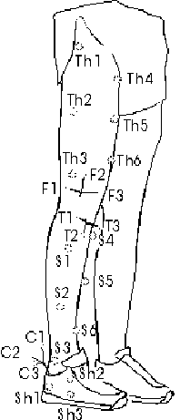
The first three thigh markers were placed at 0, 40, and 80% of the distance between the greater trochanter and the distal end of the lateral femoral condyle. Similarly, the three other thigh markers were placed at 45, 70, 95% of the distance between the anterior superior iliac spine and the proximal end of the patella. The six shank markers were also arranged on two lines. The first three shank markers were fixed at 20, 60 and 100% of the distance between the proximal end of the lateral tibial condyle and the lateral malleolus. The other three shank markers were attached at 0, 40, and 80% of the distance between the mid tibial plateau and the distal end of the tibia.
For the walking trials, the subjects wore standard running shoes (Adidas Equipment Cushioning 1994, Fig. 1) and no socks. In order to accommodate the calcaneus pin, the running shoes were slightly altered by removing the lateral part of the "Achilles tendon protector", and by removing a half-circular (r = 12 mm) piece of the heel cap at the lateral part of the right shoe.
Experimental protocol and set-up
Prior to the insertion of the pins, the subjects were given ample time to familiarize themselves with the walking procedure. The walking took place on a 10 m walkway with an embedded force plate (KISTLER, Winterthur, Switzerland). The subjects started walking approximately 5 m in front of the force plate, and were asked to hit the plate with their right feet. After finishing the stride on the force plate, the subjects continued walking for another three meters. The walking speed (1.6 0.2 m/s) was monitored with photo cells placed at equal distances (0.7 m) in front and behind the force plate (Fig. 2).
After the insertion of the pins, one standing trial and three walking trials were recorded. The standing trial was used to define the anatomical segmental coordinate systems. For the standing trial, the subjects were instructed to stand straight in a "neutral position" and to align their feet parallel to the force plate representing the laboratory coordinate system. The alignment of the subjects during the standing trials were monitored, and the subjects were asked to realign themselves if the segments (particularly the shoes) were not in alignment with the directions of the force plate. After the standing trial, the subjects were first asked to perform a few walking trials to familiarize themselves to walking with the bone pins. Per subject, three valid walking trials were recorded. Trials were discarded if the subject’s right foot was not within the force plate during the stance phase, or if the subject obviously altered his gait in order to hit the force plate.
In a pilot study, two different three-dimensional motion analysis systems were compared, a three cine-camera (LOCAM) based system and an automated video motion analysis system. After analyzing the data of the pilot study, the LOCAM based system was chosen due to better accuracy and less marker merging problems. When using 24 markers as in the present study, marker mergings occur frequently, and marker merging can be handled much better when using a manual digitizing system where the actual image is available. Even though film techniques are more accurate than video techniques, from a practical point of view, video and film techniques are comparable (Kennedy et al., 1989). Therefore, the results obtained from this study are not restricted to film techniques; the results also apply to motion recordings using current video analysis systems (or other 3D data acquisition systems) that are comparable with respect to accuracy.
The three LOCAM cameras (Fig. 2) were used at a nominal frequency of 50 Hz. Floodlights were positioned behind each camera pointing in the same direction as the camera lenses which allowed for maximum contrast between the reflective markers and the background. Prior to the motion recordings of each subject, a calibration frame with 6 control points (0.5 x 0.5 x 0.5 m3) was filmed. The calibration frame was elevated by approximately 15 cm from the floor spanning the volume from approximately marker S6 (see Fig. 1) to the mid thigh of a standing subject.
The three cameras were synchronized with the use of a threshold detector connected to the force plate (Fig. 2). The threshold detector triggered small infrared lights placed in front of the cameras, so that they were visible at the edge of the field of view of each camera. The infrared lights were active during the entire stance phase determining the duration of stance phase in each camera. Differences and fluctuations in camera speed were corrected with the use of an internal LED (200 Hz).
Three-dimensional reconstruction
For each camera and subject, the films containing the calibration frame, the standing trial and the three walking trials were digitized manually on a digitizing board (GP-8 Sonic Digitizer, SAC, Southport, CT 06490, U.S.A.). For the walking trials, two frames before and two frames after touchdown were digitized. In addition to the markers placed on the subject’s lower extremities, three stationary markers placed at the edges of the force plate were digitized in order to control for shifting of the projected film frames with respect to the digitizing board. If markers were not visible due to merging or bad contrast for less than 5 consecutive frames, the missing data points were filled with linear interpolation. Marker coordinates were normalized with respect to stance phase and the two-dimensional image coordinates were filtered with a low-pass bi-directional 4th order Butterworth filter with a cut-off frequency of 7 Hz. The cut-off frequency was determined by a residual analysis as described by Winter (1990). The appropriateness of the cut-off frequency chosen was also checked by visual inspection, i.e. by comparing the unfiltered and filtered knee and AJC rotations.
A set of programs were written in MATLAB™ (MathWorks, Natick, MA) to reconstruct the spatial positions of the markers and to calculate intersegmental motion. The spatial reconstruction was performed using a standard direct linear transformation (DLT) approach (Abdel-Aziz and Karara, 1971). For any marker at any point in time, all available cameras were used for the three-dimensional reconstruction. The program performing the spatial reconstruction always indicated which cameras were used at every instant in time for the various markers. This information allowed the monitoring and explanation of sudden changes in the spatial coordinates due to different camera combinations used for the spatial reconstruction.
The transformation of marker coordinates in a coordinate system A to the same marker in a coordinate system B was expressed as follows:
rB = [TA B] rA (1)
where: rA, rB = location vectors in reference frame (coordinate system) A and B, respectively. The location vector has the following form:
rA =
information.
Generally, the transformation matrix above can be calculated, if coordinates of 3 or more markers are known in both coordinate systems A and B. The transformation matrix represents the solution to the following least squares problem (Sderkvist and Wedin, 1993):
![]() ; n 3 (2)
; n 3 (2)
In the present investigation, a singular value decomposition method as described by Sderkvist and Wedin (1993) was employed to compute the transformation matrices. Typically, A is a local (segment-fixed) coordinate system, and B is the global coordinate system.
Reference frames and relative orientation
Kinematic data and coordinate systems were expressed similarly to what has been proposed by the ISB Standardization and Terminology Committee (Wu and Cavanagh, 1995). In the global or film coordinate system, a right handed orthogonal coordinate system was used with x-axis in the running direction, y-axis in the vertical (positive upward) direction, and z-axis in direction defined by the cross product of x and y. During the standing trial, it was arbitrarily assumed that the segmental coordinate systems were aligned with the global coordinate film system. This means that during the standing trial, the local coordinates of markers on the femur (rfem), tibia (rtib), calcaneus (rcal), thigh(rthi), shank (rsha), and shoe (rsho) were equal to the marker coordinates in the global reference (rG) frame. For any instant in time during a walking trial, the following relations existed between the segmental anatomical coordinate systems and the global coordinate system:
rG = [Tfem G] rfem (3)
rG = [Ttib G] rtib (4)
rG = [Tcal G] rcal (5)
rG = [Tthi G] rthi (6)
rG = [Tsha G] rsha (7)
rG = [Tsho G] rsho (8)
Based on these equations, the transformation matrices for the knee and AJC motion can be calculated based on bone [equations (9),(10)] and skin [equations (11),(10)] marker coordinates:
rtib = [Ttib G]-1 [Tfem G] rfem [Tfem tib] rfem (9)
rcal = [Tcal G]-1 [Ttib G] rtib [Tcal tib] rtib (10)
rsha = [Tsha G]-1 [Tthi G] rthi [Tsha thi] rthi (11)
rsho = [Tshoe G]-1 [Tsha G] rsha [Tsha sho] rsha (12)
Cardanic angles were calculated to express the rotations at the knee joint and at the AJC during the stance phase of walking based on both bone markers and skin markers. Flexion/extension, ab/adduction, and internal/external tibiofemoral rotation were resolved from the matrices using the conventions of Grood and Suntay (1983). For the ankle joint complex, plantar/dorsiflexion, ab/adduction, and in/eversion were calculated according to Cole et al. (1993). This means that for the knee rotations flexion/extension occurred around a femoral fixed medio-lateral axis, ab/adduction around the floating axis, and internal/external knee rotation around the tibial fixed proximal-distal axis. At the AJC, plantar/dorsiflexion occurred around a medio-lateral tibial fixed axis, ab/adduction around the floating axis, and in/eversion around the longitudinal (antero-posterior) foot axis.
For simplicity, knee flexion/extension can be thought to occur in a sagittal plane, ab/adduction in a frontal plane, and internal/external knee rotation in a transverse plane. Knee abduction refers to an abduction of the tibia (shank) with respect to the femur (thigh), and similarly knee adduction refers to an adduction of the tibia (shank) relative to the femur (thigh). Internal knee rotation refers to an internal rotation of the tibia (shank) with respect to the femur (thigh), and similarly, external knee rotation refers to an external rotation of the tibia (shank) relative to the femur (thigh). For the AJC, plantar/dorsiflexion can be "simplified" as the rotation of the calcaneus (shoe) relative to the tibia occurring in a sagittal plane. Similarly, ab/adduction and in/eversion of the AJC can be simplified as the relative rotations occurring in a transverse and frontal plane, respectively. For a more detailed mathematical description of the calculations involved in determining the knee and AJC rotations, the reader is referred to Grood and Suntay (1983) and Cole and co-workers (1993).
Possible differences between the external and bone marker based knee and AJC rotations are the combined effect of the skin (external marker) movement artefact occurring at the two articulating segments. In order to determine separately the contribution of the thigh and shank to knee rotation errors, the effect of the skin movement at the thigh and tibia were calculated. For these calculations, the thigh (skin) motion relative to tibia (bone) motion was determined and subtracted from the bone marker based knee rotations (femur-tibia). In this manner, the erroneous effect of the thigh could be determined. Similarly, the effect of skin markers at the shank was determined by subtracting the femur-shank based knee rotations from the femur-tibia based rotations. For the AJC rotations, the contributions of the shank and the shoe/foot segments to the difference between skin and bone marker based kinematics were determined in analogy to the analysis of the tibiofemoral motion as described above.
Motion recordings of bone movement with the use of intracortical pins is a highly invasive procedure. It may be argued that the insertion of the pins may cause discomfort and the anesthetics may alter the subject’s perception. For two of the subjects (subject 2 and 4), a dry run with exactly the same motion recordings and skin markers was performed prior to surgery, in order to show quantitatively whether the subject’s gait was affected due to the insertion of the bone pins.
For the present study, it was assumed that the skin marker movement was not restricted by the insertion of the pins. In order to minimize this effect, skin markers were placed at least 5 cm away from the closest bone pin insertion. By manually moving the skin markers closest to the pin insertion, it appeared that the skin movement was not restricted at these locations due to the bone pin.
The calibration frame available at the site of the experiments was limited due to the few calibration points and its size. The accuracy of spatial reconstruction is reduced when a small number of calibration points are used (Hatze, 1988) or when markers are reconstructed that move outside the calibration volume (Wood and Marshall, 1986). Since the upper thigh markers moved outside the calibration volume, some errors may be introduced when using these markers to calculate skin marker based tibiofemoral joint motion. The magnitude and effect of these errors were determined by comparing joint motions based on all skin markers and the skin markers closest to the bone pins. Additionally, the residuals of the spatial reconstruction were calculated for each marker indicating the appropriateness of the DLT model for each of the skin and bone markers.
Problems were encountered with the femur pin in two subjects. In one subject (subject 2), the femur pin became loose after a few walking trials. The femur pin was then surgically removed and the experiments were continued with the remaining markers. Consequently, data of this subject will only be available to describe the AJC motion. It was suspected that the slit cut into the iliotibial band through which the femur pin was inserted may have been too small in subject 2. This may have caused large forces between the femur pin and the iliotibial band, eventually resulting in the loosening of the pin. In another subject (subject 4), it was observed that a "popping", a sudden rotation, of the femur pin occurred. This "popping" movement of the pin was in the order of 10o flexion/extension, and appeared to occur in both directions during the swing phase when the knee underwent relatively large flexion angles. This popping movement did not represent true motion of the femur, since this sudden flexion movement would correspond to a large relative translation between the femoral head and the pelvis. Therefore, it was decided to discard the femur pin data for this subject. Consequently, tibiofemoral kinematics will only be presented for three subjects (subject 1, 3, and 5).
None of the subjects experienced pain and/or significant discomfort during the walking and running trials (chapters 4 to 6) used in this study. However, one subject did experience minor pain and discomfort in the distal part of the calcaneus during the later part of the experiments (barefoot running trials) which were not part of this project (see Fig. 17). During the motion trials, none of the subjects stated that his ability to walk or run normally (chapters 4 to 6) was affected by the pins
Qualitatively, the gait of all the subjects appeared to be the same with bone pin compared to the gait exhibited during the training sessions (used to familiarize the subjects with the walking and running (see chapters 4 to 6) procedure). The qualitative assessment of the subject’s gait was confirmed by comparing the skin marker based kinematics for walking with and without the bone pins. At the knee joint, the difference in both subjects at any instant during the stance phase did not exceed 2.1 for ab/adduction, 4.8 for internal/external knee rotation and 4.5 for flexion/extension. At the AJC, the absolute differences did not exceed 6.0 for in/eversion, 4.8 for ab/adduction and 4.8 for plantar/dorsiflexion.
These differences may be considered to be substantial, but the amplitude and shape of the curves were similar; the major part of the difference in skin based joint kinematics between the pin trials and the non-pin trials was a systematic shift between the two curves (Fig. 3). This systematic shift can be attributed to the different standing trials used for the pin and the non-pin walking trials. Knee rotations based on skin markers for the pin and the non-pin trials are displayed for one subject (subject 2) in Fig. 3. The trends were similar for the other subject and for the AJC motions, i.e. the shape and amplitude of the skin and bone marker based curves were similar.
Accuracy of spatial reconstruction
Residuals |
Sub. 1 |
Sub. 3 |
Sub. 5 |
Femur Markers (F1 to F3) Thigh Markers (Th2 to Th6) |
11.5 17.1 |
10.0 12.0 |
13.9 13.9 |
The residuals of the DLT calculations are plotted in Fig. 4 for subjects 1, 3, and 5. The (skin) markers at the thigh were of primary interest since these markers were clearly outside the volume calibrated with the frame. The results (Fig. 4) show that only the residuals for thigh marker 1 (Th1, see Fig. 1) placed over the greater trochanter appeared to be consistently higher than the values of the other thigh markers (as well as the femur markers). This marker was only seen at the edge of the film in all three cameras where errors due to lens distortion can be expected to be high. This marker was excluded for the calculations of the knee motions even though the difference in knee rotations between calculations including and excluding this marker was found to be systematic and small (<2.5 for flexion, and <1.5 for internal/external rotation and ab/adduction).
The residual plots of the various markers also show that the residuals were generally higher at the beginning and end of the stance phase (Fig. 4). This result was not surprising. The synchronization method used allowed synchronization of the cameras to within one film frame (50 Hz corresponding to 20 ms). Therefore, one camera records the marker at t1 and another camera records the marker at time t1 +
Dt where Dt can be as high as 20 ms. The effect of this "asynchronicity" of the cameras can be expected to be higher during phases when fast movements occur such as during the touchdown and takeoff phases. This explains why the residuals of the various bone and external markers were generally higher at the beginning and end of the stance phase of walking.Differences between skin and bone marker based tibiofemoral rotations may potentially be masked by inaccuracies in the DLT calculations for the markers moving outside the calibration volume, i.e. the thigh markers. In order to estimate this error source, the following calculations were made. First, the average residuals were calculated from Fig. 4 for all the femur and thigh markers (excluding the marker at the greater trochanter, Th1) during the stance phase. These values are displayed in Table 2. Taking the worst case (subject 1), the difference between femur and thigh markers was 5.6 (subject 1) which corresponds to 1.7 mm. Assuming an average marker distance of 150 mm, 1.7 mm uncertainty corresponds to roughly 0.6 (= arctan ) error in rotation. However, it should be realized this error is likely to be much smaller since the markers outside the calibration volume would be moved (distorted) in a similar, systematic manner which would result in less than the estimated 0.6 error in rotation. Based on these calculations, differences between external and skeletal marker based rotations exceeding 0.6 can not be explained by inaccuracies due to the DLT calculations.
Due to technical problems (overexposure, film frequency slowing down) in some subjects all three cameras were not available for the first or the last part of the stance phase of the walking trials. During these times, cameras 1 and 2 were typically the only cameras available. Since the angle between camera 1 and 2 was rather small (35, see Fig. 2), the spatial position of the markers can be expected to be less accurate. The transition from three to two cameras (and vice versa) typically caused "jumps" in the curves. Therefore, the times where only two cameras were available were marked in the following graphs (Fig. 5, Fig. 6), and all subsequent calculations (Table 3, Table 4) did not include these phases where only two cameras were available.
The individual knee rotations derived from skin and bone markers are depicted in Fig. 5. Additionally, average differences (root mean square, RMS), maximal differences and general curve agreement between tibiofemoral rotations derived from skin and bone markers are presented in Table 3. Generally, the difference in skin and bone marker based knee rotations between trials and within subjects was small (Fig. 5), i.e. the knee kinematics were very repeatable. The intersubject variability was typically much larger than the intrasubject variability (see Fig. 5).
The bone marker based knee ab/adduction movement patterns were different for the three subjects analyzed, i.e. no general ab/adduction pattern could be observed across subjects (Fig. 5). The ab/adduction range of motion (ROM) also varied across subjects; the ROM was about 5 for subjects 1 and 3, whereas the range of motion in ab/adduction was around 10 for subject 5. The difference between skin and bone marker based ab/adduction varied across subjects (Fig. 5). The best agreement was found in subject 1 where the difference between skin and bone marker based curves did not exceed 3.1 (Table 3). Additionally, the shape of the skin and bone marker based average curves was similar for subject 1, whereas in subject 3 and 5 poor agreement was found between the shape of the skin and bone marker based curves. The maximum difference between ab/adduction calculated from skin and bone markers was present in subject 5 where the maximal difference exceeded 5 (Table 3).
The patterns of internal/external knee rotation derived from bone markers varied across subjects, e.g. a pronounced initial internal tibial rotation with respect to the femur was present in subject 3, whereas for the other two subjects, no (subject 5) or only a small amount (subject 1) of initial internal knee rotation was observed (Fig. 5). The range of motion of internal/external (longitudinal) knee rotation derived from bone markers was around 5 for subject 1 and 5, whereas for subject 3, the range of motion was more than 10. Poor or virtually no agreement between skin and bone marker based kinematics was found in subject 1 and 5. On the other hand, relatively good agreement was present in subject 3, in particular for the time from 25% of stance phase to take-off. In all three subjects, the skin marker based internal/external rotation showed a distinct initial internal tibial rotation with respect to the femur, which was either not present at all or only to a much lesser extent in the bone motion. For all subjects, the maximal difference between knee rotation derived from skin and bone markers exceeded 7 (Table 3).
Knee Rotation |
Variable |
Sub. 1 |
Sub. 3 |
Sub. 5 |
Mean |
Ad/ Abduction |
RMS Diff. [ ]Max. Diff. [ ] |
2.1 3.1 |
2.4 4.0 |
2.8 6.0 |
2.4 4.4 |
Shape Agreement |
good |
poor |
poor |
poor |
|
Int./ Ext. Rotation |
RMS Diff. [ ]Max. Diff. [ ] |
4.2 7.6 |
2.1 7.3 |
5.3 10.3 |
3.9 8.4 |
Shape Agreement |
poor |
good |
poor |
poor |
|
Flexion/ Extension |
RMS Diff. [ ]Max. Diff. [ ] |
1.5 2.6 |
1.7 4.6 |
3.2 5.8 |
2.1 4.3 |
Shape Agreement |
excellent |
excellent |
good |
excellent |
The shape of knee flexion/extension based on bone markers was similar for all subjects. The amplitude of the movement of flexion/extension, however, varied between subjects. Subjects 1 and 3 had much more flexion/extension movement from touchdown throughout the end of midstance. Generally, the agreement between bone and skin marker based knee flexion was good, especially during the first half of stance phase. The least agreement between flexion/extension derived from skin and bone markers was present in subject 5, where the maximum difference was as high as 5.8 (Table 3).
The individual AJC rotations derived from skin and bone markers are presented in Fig. 6, and for each subject, average (RMS) and maximal differences between skin and bone marker based rotations were calculated (Table 4). Similarly to the knee motions, the intrasubject differences between trials were small compared to the intersubject variability.
AJC Rot. |
Variable |
Sub. 1 |
Sub. 2 |
Sub. 3 |
Sub. 4 |
Sub. 5 |
Mean |
In/ Eversion |
RMS Diff. [ ]Max. Diff. [ ] |
4.4 6.4 |
3.4 5.3 |
3.6 5.6 |
2.5 7.1 |
2.9 4.2 |
3.4 5.7 |
Shape Agreement |
good |
good |
good |
good |
good |
good |
|
Ad/ Abduction |
RMS Diff. [ ]Max. Diff. [ ] |
4.3 5.7 |
1.4 2.5 |
2.0 5.0 |
1.4 3.1 |
3.2 4.5 |
2.5 4.2 |
Shape Agreement |
good |
excellent |
good |
excellent |
good |
good |
|
Plantar./ Dorsiflexion |
RMS Diff. [ ]Max. Diff. [ ] |
3.1 4.9 |
2.2 5.8 |
4.4 8.1 |
3.1 5.8 |
2.5 4.6 |
3.1 5.8 |
Shape Agreement |
good |
excellent |
good |
excellent |
excellent |
excellent |
The shape of the bone marker based in/eversion curve was similar across all five subjects. An initial eversion movement was typically followed by an inversion movement towards the end of stance phase. The initial eversion calculated from the bone markers as the difference between the eversion at touchdown and maximal eversion (during the first 50% of stance) was in the same order of magnitude (5) for all subjects. On the other hand, initial eversion calculated from skin markers was much higher, and ranged between 10 and 16 depending on the subject. In all subjects, the eversion indicated by the skin markers was much higher than the eversion that actually took place between tibia and calcaneus. The difference between skin/shoe and bone marker based in/eversion was highest from around 20 to 60% of stance phase, and was in the order of magnitude of 5, a substantial difference considering the amplitude of this motion.
The tibiocalcaneal ab/adduction patterns of the bone motion were similar across subjects. An initial abduction during the first 20% of stance was generally followed by a slow adduction movement. The ab/adduction exhibited at the AJC was generally well reflected with the use of skin/shoe markers, in particular for three subjects (subjects 2, 3, and 4). For subject 1 and 5 more abduction was indicated with the skin/shoe markers. However, the shape of the average curves for skin and bone marker based kinematics were similar.
The shape of the plantar/dorsiflexion derived from bone markers was similar across subjects. Typically, the plantar/dorsiflexion curves had two peaks. The first plantarflexion peak occurred at around 10% of stance phase, whereas the maximum dorsiflexion movement typically occurred at 75% of stance phase. The difference between skin and bone marker based kinematics appeared to be systematic, in the sense that the amplitude of the movement was always higher for the skin marker based kinematics. That means that the maximum plantarflexion as well as the maximum dorsiflexion were overestimated when using skin markers. The overestimation of plantarflexion varied between subjects and could be as high as 5.8 as seen in subject 4 (Table 4). The overestimation of the maximum dorsiflexion movement which occurred around 75% of stance phase ranged from 1 (subject 2) to 8 (subject 3).
The rotational knee motions found in this study were generally in agreement with the tibiofemoral rotational kinematics reported in previous investigations (Lafortune et al., 1992a; Lafortune et al., 1994). The knee flexion/extension curves were similar in shape and magnitude to the data presented by Lafortune et al. (1992a). The range of motion in ab/adduction was much higher than the range of motion reported by Lafortune et al. (1992a). Their data indicated that almost no ab/adduction movement took place during stance. The difference between their results and the results of this study may be explained by differences in defining the anatomical coordinate systems of the tibia and femur (see chapter 6). Lafortune et al. (1992a) employed anatomical coordinate systems based on a roentgen-stereophotogrammetric analysis, whereas in the present study the anatomical coordinate systems were based on a "neutral" standing trial. It also appeared that the ab/adduction range measured for subject 5 was unphysiologically high. This may likely be the result of an alignment problem of the anatomical coordinate system causing cross-talk from the knee flexion/extension (see chapter 6).
In the study of Lafortune et al. (1992a), all five subjects clearly showed an initial internal tibial rotation with respect to the femur (internal knee rotation). Based on the curves presented by Lafortune and co-workers (1992a), the initial internal knee rotation was estimated to range from 2 to 6.3 across subjects. The results of this study showed minimal initial internal knee rotation of less than 2 for subject 1, a clear initial internal knee rotation of 5 for subject 3, and initial external knee rotation of approximately 4 for subject 5 (Fig. 5). The findings of this study show, therefore, more intersubject variability than the findings of Lafortune et al. (1992a). In order to discuss the results of the present investigation, the internal/external tibial and femoral rotations were also calculated with respect to the global laboratory coordinate system. The results showed that the tibia rotated internally from touchdown to about 25% of stance phase. This initial tibial rotation with respect to the global coordinate system was consistently present in all trials except in one trial of subject 3 where the tibia showed a short initial external rotation of less than 2 changing into internal tibial rotation of 4.4 after 7% of stance phase. The initial tibial rotation with respect to the laboratory coordinate system averaged 10.1, 5.6, and 8.9 for subject 1,3, and 5, respectively. This initial rotation was also present at the femur and averaged 8.6 (subject 1), 4.2 (subject 3), and 11.0 (subject 5). These results support the generally accepted paradigm of internal tibial rotation at and shortly after touch-down. However, this internal tibial rotation appears to be matched by internal femoral rotation. In subject 5, it was actually found that this internal rotation was larger at the femur which resulted in an external knee rotation.
Based on the results of this study, it can be concluded that skin markers as used in this study can only be used to reliably (maximal error < 20% of range of motion) determine flexion/extension at the tibiofemoral joint. For knee flexion/extension, the differences between bone and skin marker based knee kinematics was not only smallest in absolute terms, but especially in relation to the amplitude of the movement (signal to noise ratio). For knee ab/adduction and internal/external rotation, the error introduced as a result of the skin movement artefact can be almost as high as the motion measured. Therefore, knee ab/adduction and internal/external rotation calculated from skin markers as used in this study has to be interpreted with extreme caution in particular when differences across subjects are of interest. It may be argued that large differences within the same subject, e.g. a pre-intervention to a post-intervention comparison of a patient, may be detected in all knee rotations with the use of skin markers. However, the authors are convinced that such comparisons are not appropriate and suggest that the interpretations of such results may lead to wrong conclusions since the difference seen in the skin marker based kinematics may not reflect the true differences at the tibiofemoral joint. Statements other than that "there are differences" should be considered as overinterpretations. Additionally, the inability to reproduce the same anatomical coordinate system even within subjects may also cause or mask possible differences (see also chapter 6).
The discrepancies between skin and bone marker based kinematics for the knee joint (Fig. 5) are the combined effect of the skin movement artefact at the thigh and shank. Subject averages of the errors in knee rotations caused by the skin movement artefact at the thigh and tibia are depicted in Fig. 7. The curves show that most of the knee rotation errors occurred due to skin movement artefacts at the thigh. Shank induced errors were small, especially for knee ab/adduction (<2.6) and knee flexion/extension (<1.8). The magnitude of the errors due to skin movement artefact at the shank were in agreement with results from a previous investigation (Holden et al., 1994). Additionally, the results of Holden and co-workers (1994) also showed that the largest difference between rotational movement of the shank as determined from skin markers and tibia as determined from bone markers existed along the longitudinal axis of the lower leg. Based on the results of this investigation, it may be argued that the skin movement artefact at the shank can be neglected in comparison to the errors caused by the thigh. The result that thigh induced errors were much higher was expected. The thigh consists of much more soft tissue (muscles, adipose tissue) than the shank, and therefore, relative movement between skin and underlying bone was expected to be much higher than for the leg. Furthermore, the adherence or attachment of the soft tissue to the underlying bone may be different for the thigh and the shank. For example, the soft tissues over the femoral condyles may be rather thin but loose. On the other hand, the soft tissue over the tibia crest may be equally thin but relatively firmly attached to the bone.
No studies are known to the authors that investigated AJC (tibiocalcaneal) motion by actually measuring the motion of the respective bones during gait. Hence, gait analysis studies using external markers have to be used to compare the results of this study. Generally, the movement patterns of the skin marker based AJC motion was in agreement to what has been reported by Moseley and co-workers (1996). However, in the present study, the ranges of the AJC rotations were higher than the respective ranges presented by Moseley et al. (1996). This difference may be attributed to the fact the subjects in this study wore shoes, whereas in the other study the AJC rotations were determined when walking barefoot.
Unlike the rotations measured at the knee joint, the rotations at the AJC were more uniform across subjects. In general, the shape of the rotation curves were similar for all subjects, the only difference consisted in the magnitude, i.e. in the range of motion.
In absolute terms, the smallest difference between skin and bone marker based AJC motion was measured for ab/adduction. For subjects 2, 3 and 4, ab/adduction determined from skin and bone markers were almost identical. For subject 1 and 5, ab/adduction was overestimated by about 5. Hence, it can be concluded that ab/adduction patterns of the calcaneus with respect to the tibia can be determined reliably when using external (skin) markers as used in this investigation. For in/eversion, the skin and bone marker based results showed that the skin marker based in/eversion had similar patterns as the bone marker based in/eversion, however, with a higher amplitude. Similarly, the bone marker based plantar/dorsiflexion was well reflected by the skin marker based result, but again with a higher amplitude in both plantarflexion and dorsiflexion. The reason for the phenomenon that the calcaneus-tibia rotation were amplified when measured with external markers may be twofold. Firstly, the movement of the shoe may be different from the movement of the foot (Stacoff et al., 1992; Reinschmidt et al., 1992). This will be discussed in more detail later. Secondly, some of the movement between foot/shoe and shank may occur in the talonavicular joint rather than in the joints (talocalcaneal and talocrural joint) located between calcaneus and tibia.
The contribution of the shank and the shoe/foot segment to the difference between skin and bone marker based AJC kinematics was determined in analogy to the analysis for the tibiofemoral joint and is displayed in Fig. 8. Similar to the tibiofemoral joint, the least amount of error was introduced by the shank. The error due to the skin movement artefact at the shank was in the range of 2-3 and only exceeded 5 in in/eversion for one particular subject. The misreadings caused by the relative movement of the shoe markers with respect to the calcaneus were as high as 7, and were in the average always higher than the errors due to skin movement artefact at the shank. The error patterns caused by the shoe/foot segment were similar across all the subjects in all three AJC rotations. On the other hand, there were no apparent error patterns in the AJC kinematics for the errors caused by the shank. This observation was similar to what could be observed for the errors due to the shank at the tibiofemoral joint where again no apparent error patterns for the shank were found. On the other hand at the tibiofemoral joint, the error patterns caused by the thigh were somewhat similar across subjects (Fig. 7), but they were not as uniform across subjects as the error patterns caused by the shoe/foot at the AJC (Fig. 8). This observation implies that the skin movement artefact is more predictable for the AJC than for the knee joint. However, it has to be realized that the similarity in error patterns at the knee and AJC may be dependent on the homogeneity of the subjects analyzed. The subjects included in this study were rather homogenous. None of the subjects could be considered obese, they were all male, and they were all about the same age. The challenge of developing a model to correct for skin movement artefact for any subject would be to account for individual factors such as the amount of adipose tissue in the lower extremities and the movability of the skin with respect to the underlying tissue. This has also been pointed out by Holden and co-workers (1994) who suggested that a generalized model of soft tissue error (at the shank) has to account for individual subject differences.
The relative motion of skin with respect to the underlying bone can be caused by inertial effects, the non-rigid attachment of the skin to the bone, and by movement caused by muscle contractions underneath the skin. Artefacts due to inertial effects should primarily occur during the first 50 ms of stance phase, the impact phase, and should be minimal during the remaining stance and swing phase. Artefacts due to muscle activity can occur during the entire stance and swing phase. It is difficult to estimate the contribution of these effects on the relative movement between skin and underlying bone. Based on the qualitative observation of the films, it was felt that the skin movement artefact during the stance phase was mainly caused by muscle movements. Qualitative assessment also suggested that the skin artefacts due to muscle movements may even be higher during the swing phase than during the stance phase analyzed in this investigation. Another problem connected with movement due to muscle contraction is the standing trials being used to determine the anatomical position of the segments. During the standing trial, muscle can be expected to be in a state of low activity. However, during the stance phase of walking some muscles are always activated. Therefore, part of the skin movement artefact may simply be caused by the fact that standing trials were usually recorded with a different muscle activity as occurring during walking.
The skin movement artefacts at the shoe/foot segment have to be considered differently than the skin movement artefact occurring at the shank or tibia. A shell (shoe) is added on top of the skin which may introduce an additional relative motion. Therefore, the movement between shoe and calcaneus can be broken down into the relative movement between the shoe and foot and the movement between foot and calcaneus. Based on the data gathered in this study, it cannot be concluded if the skin movement artefact seen at the AJC was mainly due to the relative movement between the shoe and foot or due to the relative movement between foot and calcaneus. However, based on the fact that part of the heel counter had to be removed to accommodate the bone pin may suggest that the heel counters of these shoes were not very firm. This may have allowed for a considerable relative motion between the shoe and foot. The result that the movements based on the shoe markers were generally larger than the actual movement was in the same direction as the differences found in investigations comparing the rearfoot movement based on shoe and foot markers as viewed through windows cut into the shoe (Stacoff et al., 1992; Reinschmidt et al., 1992). It may be argued that the skin movement artefact caused by the relative motion between shoe and calcaneus was mainly caused by the relative motion between shoe and heel and not between heel and calcaneus. In other words, if windows were cut into the shoe and markers were attached to the skin of the heel, the tibiocalcaneal motion may have been better estimated than with the markers attached to the shoe.
The results of this study showed that tibiofemoral rotations determined with external markers as employed in this study have to be used and interpreted with caution for any type of gait analysis. Knee rotations other than flexion/extension are small and the errors induced through skin movement artefacts may well exceed the actual motion occurring at the tibiofemoral joint. For some subjects, there was no agreement in shape of ab/adduction or internal/external knee rotations as determined with skin and bone markers. Therefore, it is suggested that knee rotations other than flexion/extension cannot be reliably determined when using external skin markers. The segmental error analysis revealed that the discrepancies between skin and bone marker based kinematics were almost exclusively caused by the skin movement artefact occurring at the thigh. Therefore, external markers may still be used to measure the rotations of the tibia with respect to a laboratory coordinate system. This will be discussed in detail in chapter 7.
At the ankle joint complex, skin/shoe marker based kinematics gave a relatively good estimate of the actual tibiocalcaneal motion. In particular, the shape of in/eversion, ab/adduction, and plantar/dorsiflexion at the AJC was well reflected with the use of external markers. However, the AJC rotations are generally overestimated when using external markers. Therefore, skin/shoe markers may be used to "reflect" the AJC motion, however, absolute values have to be used and interpreted with caution.
Skin mounted markers are typically employed in kinematic gait analysis. However, large errors may be introduced as a result of the relative movement between skin and underlying bone. To date, knowledge about this skin movement artefact is limited, and no studies have been published which systematically investigated this error at the thigh, shank, and foot/shoe during a walking movement. The purpose of this study was to determine the errors in knee (tibiofemoral) and ankle joint complex, AJC, (tibiocalcaneal) rotations caused by the skin movement artefact during walking. Intracortical bone pins were inserted into the femur, tibia, and calcaneus of five subjects. Marker triads were attached to these pins, and additionally, six skin markers to the thigh, six to the shank, and three to the shoe. For each subject, three walking trials were filmed with three synchronized LOCAM cameras (50 Hz) to determine the 3-D position of the markers during stance phase. Flexion/extension, ab/adduction, and longitudinal rotation at the tibiofemoral joint as well as plantar/dorsiflexion, ab/adduction, and in/eversion at the AJC were calculated from both skin and bone markers. The results showed that the errors in knee rotations were mainly caused by the thigh markers. Knee flexion/extension was generally well reflected with the use of skin markers (mean difference 2.1). Depending on the subject, the agreement between skin and bone marker based kinematics for ab/adduction and internal/external knee rotation ranged from good to virtually no agreement, and in some subjects, the errors exceeded the actual motion. The errors in AJC rotations were mainly caused by the markers on the shoe/foot segment. The tibiocalcaneal motions were generally well reflected with external markers. However, tibiocalcaneal rotations derived from external markers typically exceeded the true bone motions. The results suggest that (a) knee rotations other than flexion/extension may be affected with substantial errors when using external markers, and (b) tibiocalcaneal motions are generally well reflected with external markers, but absolute values (amplitudes) have to be interpreted with caution.
CHAPTER 4:
Tibiocalcaneal Motion During Running - Measured With External And Bone Markers
To date, numerous studies have investigated the kinematics of the lower extremities and in particular the kinematics of the rearfoot with respect to the lower leg during running. These studies have been conducted for a number of reasons such as to gain a better understanding of the joint kinematics during running (Engsberg and Andrews, 1987), to determine the influence of running shoe parameters (e.g. Nigg and Morlock, 1987) or foot morpholgy (Nigg et al., 1993) on running kinematics, to investigate kinematic changes due to orthotics (e.g. Taunton et al., 1985), or to study a possible relationship between kinematic parameters and running injuries (e.g. Messier and Pittala, 1988). One of the main interest of these studies was to quantify the movement of the calcaneus with respect to the tibia. To approximate these bony movements, markers or linkages were typically attached to the leg as well as to the foot or the shoe. However, during a highly dynamic movement such as running, these external markers or linkages may move considerably with respect to the underlying bone. Therefore, a large error, typically referred to as the skin or shoe movement artefact, may be introduced as a result of this relative movement.
This skin movement artefact has motivated researchers to directly measure in-vivo skeletal motion during human ambulation (Levens et al., 1948; Karlsson, 1990; McClay, 1990; Murphy, 1990; Lafortune et al., 1992a; Cappozzo et al., 1996; Holden et al., 1994; Lafortune et al., 1994) even though procedures to directly measure in-vivo skeletal motion are typically highly invasive. Most of these studies were confined to slow movements such as walking, and to date, only one study determined the skeletal (patellofemoral, tibiofemoral) motion during running (McClay, 1990). However, no study has been conducted that determined the skeletal tibiocalcaneal motion during running.
The effect of the skin movement artefact on kinematics of the lower extremities has been determined in a few investigations looking at slow or quasi-static movements (Lafortune et al., 1992b; Cappozzo et al., 1996; Maslen and Ackland, 1994; Karlsson and Lundberg, 1994). Generally, large discrepancies were reported between the external marker and skeletal marker based motions during these slow movements. The effect of the skin movement artefact is highly dependent on the joint or joint complex in consideration. In chapter 3, it was shown that during walking tibiocalcaneal rotations (in/eversion, ab/adduction, plantar/dorsiflexion) were fairly well represented when using external markers; but, for tibiofemoral rotations, only flexion/extension was well represented when using external markers. In this context, it is of interest if external markers can also be used reliably to determine tibiocalcaneal motion during a more dynamic movement such as running.
Rearfoot (tibiocalcaneal) kinematics and in particular in/eversion of the rearfoot is believed to be important with respect to running injuries and the design of running shoes. However to date, it has not been shown how much rotation actually occurs between the calcaneus and tibia during running, and how accurately tibiocalcaneal rotations can be measured or estimated when using external markers. Therefore, the purpose of this investigation was (a) to determine tibiocalcaneal rotations during running as derived from bone markers, and (b) to compare the skeletal tibiocalcaneal rotations with rotations as derived from external markers attached to the shank and the shoe.
Methods used in this study were similar to the one presented in chapter 3, and therefore, the methods will only be described briefly. The main difference to the methods presented in the previous chapter are that (a) running (speed 2.9 m/s) kinematics were assessed, (b) the motion was recorded at 200 Hz, (c) five trials were collected per subject, (d) only the results for the AJC rotations are reported, and (e) a typical variable used in kinematic assessments of running, the maximal initial eversion, was calculated based on external as well as skeletal markers. The reader who is already familiar with the method section of the previous chapter may want to move directly to the results section of this chapter.
Five male subjects (age 28.6 4.3 yrs., weight 83.4 10.2 kg, height 185.1 4.5 cm) gave informed consent to participate in this study. The subjects had neither an injury at the time of testing nor an injury history which may have possibly lead to an abnormal running style. The experimental protocol was approved both by the ethics committees of The University of Calgary, Canada, and the Karolinska Hospital in Stockholm, Sweden. The experiments were conducted at the Department of Orthopaedics of the University Hospital in Huddinge, Sweden.
Intracortical Hofmann bone pins (2.5 mm diameter) were inserted into the posterolateral aspect of the calcaneus and into the lateral tibial condyle (Gerdy’s tubercle) in the subject’s right leg. Standard local anesthetic (Citanest 10 mg/ml) was applied at the insertion sites of the bone pins. To each of the bone pins, triads of reflective markers (10 mm diameter spheres) were attached. In addition to these bone pins, external markers were attached to the segments of interest: six external reflective markers (20 mm diameter spheres) were fixed to the skin of the shank at standardized locations, and three external markers were glued directly to the posterior aspect of the shoe (Fig. 1). The subjects wore standard running shoes (Adidas Equipment Cushioning 1994, Fig. 1), where a small half-circular piece (r = 12 mm) of the lateral heel cap (of the right shoe) was removed in order to accommodate the calcaneus pin. The subjects were barefoot in these shoes (they did not wear socks).
Protocol and motion recordings
For each subject, one standing trial and five heel-toe running trials (2.9 0.2 m/s) were collected. Prior to the running trials and surgery, the subjects had ample time to familiarize themselves with the running procedure. The subjects were asked to land on a force plate (Kistler, Winterthur, Switzerland) with their right shoe. The force plate was embedded in the floor of a 10 m runway, and the speed was monitored with photo cells placed 70 cm in front and behind the force plate. The marker motions were recorded during the stance phase with the use of three high speed cine cameras (LOCAM) operating at a nominal frequency of 200 Hz. The cameras were placed at 10, 55 and 110 to the running direction (Fig. 2). The cameras were synchronized through small infrared lights visible in all cameras. The lights indicated the stance phase and they were triggered with a threshold detector connected to the force plate. Differences and fluctuations in camera speed were corrected using an internal LED producing small dots at the side of the film in 5 ms intervals. At the beginning of all trials, a high precision calibration frame with 6 control points was filmed to allow the three-dimensional reconstruction.
In addition to the five bone pin trials, two subjects (subjects 2 and 4) also performed three "pre-operative" runs and a standing trial prior to inserting the bone pins. For these pre-operative runs, the external markers were placed at the same locations as for the bone pin trials. The comparison of the tibiocalcaneal rotations based on external markers with and without bone pins was used to determine whether the insertion of the bone pins affected the "natural" running style of the subjects.
The films of the calibration, standing and running trials of each subject and camera were projected onto a digitizing board (GP-8, Sonic Digitizer, SAC) and manually digitized. Reflective markers at the edge of the force plate were used to control for shifting of the projected image with respect to the digitizing board. For filtering purposes, five frames before and five frames after stance phase were analyzed for each trial of each camera. If markers were not visible for less than five consecutive frames due to poor contrast or marker merging, the missing coordinates were linearly interpolated. Marker coordinates were normalized to 101 data points with respect to stance phase with 0% being touchdown and 100% being take-off. Camera coordinates were filtered with a bi-directional 4th order low-pass Butterworth filter with a cut-off frequency of 10 Hz determined from a residual analysis (Winter, 1990).
A standard direct linear transformation (DLT) using 11 coefficients (Abdel-Aziz and Karara, 1971) was used to calculate the spatial position of all the markers during the stance phase of running as well as during the standing trial. The anatomical segmental reference frames for the calcaneus, tibia, shoe, and shank were defined by using the standing trial where the subjects were instructed to align their lower limb segments with the global (film) reference frame. Coordinate transformations were applied to calculate the transformation matrix transforming the anatomical tibia (shank) reference frame into the anatomical calcaneus (shoe) reference frame. A singular value decomposition method (Sderkvist and Wedin, 1993) was employed to calculate the respective transformation matrices. For the transformation matrices concerning the shank, all skin markers were used that were visible in all the cameras during all frames of the stance phase. That means that depending on the subject, four to six shank markers were used for these calculations.
Intersegmental tibiocalcaneal motion which can be viewed as the combined motion of talocrural and talocalcaneal motion, was expressed by Cardanic angles using the sequences as proposed by Cole et al. (1993). That means that for the bone marker based rotations, plantar/dorsiflexion was calculated around the medio-lateral tibial fixed axis, ab/adduction around the floating axis, and in/eversion around the antero-posterior calcaneus axis. Similarly for the skin marker based tibiocalcaneal kinematics, plantar/dorsiflexion was resolved around the medio-lateral shank fixed axis, ab/adduction around the floating axis, and in/eversion around the antero-posterior shoe axis. Note that the terms shank and shoe will be used to refer to a body segment defined by external (surface) markers, and tibia and calcaneus will refer to the corresponding skeletal segments. The procedures involved with calculating the coordinate transformations and intersegmental motions were already described in detail in chapter 3.
A segmental error analysis was performed in order to determine the contribution of each segment to the difference in intersegmental rotations as derived from external and bone markers. For these calculations, the rotations of the shank with respect to the calcaneus were calculated and compared to the skeletal tibiocalcaneal rotations. Similarly, the rotations of the shoe with respect to the tibia was subtracted from the tibiocalcaneal rotations in order to determine the error caused by the relative movement of the shoe markers with respect to the underlying calcaneus.
The differences between bone and skin marker based tibiocalcaneal rotations (plantar/dorsiflexion, ab/adduction, in/eversion) were expressed in terms of the average (of single trials) root mean square (RMS) of the difference between the three rotation angles derived from bone and external markers at every instant in time during the stance phase of running. Additionally, the maximal RMS difference was calculated for each of the subjects during the stance phase of running.
In order to compare the effect of possible differences between bone and external markers based kinematics, the maximal change in eversion (
Dbmaxext, Dbmaxbone), a variable typically used in studies concerning rearfoot kinematics, was determined for eversion derived from both external and bone markers. The maximal change in eversion was calculated as the change in eversion from touchdown to the maximal value of eversion. A two-way analysis of variance (ANOVA) with a complete block design (subject) was employed to detect significant differences in maximal eversion derived from external markers and bone markers. The level of significance was set at P < 0.05.
Effect of pins on running kinematics
Tibiocalcaneal rotations based on external markers before and after inserting the bone pins were determined for subject 2 and 4 (Fig. 9). In almost all of the rotations, a systematic shift was present between the two average curves (Fig. 9). This systematic shift was attributed to different standing trials used for the pin and non-pin running trials (see also chapter 3, page *). When correcting for the systematic shift, the difference in skin marker based tibiocalcaneal rotations between the pre-operative runs and the bone pin trials was highest for the ab/adduction in subject 2 (3.6). However, this difference was mainly caused by an "outlier" trial which increased the average abduction calculated from the external markers of the bone pin trials (Fig. 9). In general, the graphs (Fig. 9) show that the shape and magnitude of the skin marker based rotations measured with and without bone pins were very similar. Therefore, it was concluded that the subjects did not change (or only minimally changed) their running style due to psychological and/or proprioceptive effects caused by the bone pins.
The tibiocalcaneal rotations are presented in Fig. 10. In/eversion patterns were consistent across subjects. The position at touchdown ranged from neutral (0) to 5 inversion. In all subjects, an initial eversion took place reaching a maximal value at around 50% of stance phase. After the point of maximal eversion, the calcaneus continually inverted with respect to the tibia. Typically, this inversion motion was slightly higher than the initial eversion motion. At take-off, the calcaneus (relative to the tibia) was in an inverted position in all subjects and trials. The in/eversion excursion during the stance phase ranged between 10 (subject 3) and 17 (subject 1).
The ab/adduction patterns were similar across subjects, however, it appeared that the inter-subject variability in ab/adduction was greater than the inter-subject variability in/eversion and plantar/dorsiflexion. The range of motion in ab/adduction was small (average 7.5) compared to the other tibiocalcaneal rotations. All subjects showed an initial abduction followed by a larger adduction towards the end of stance phase.
All subjects landed in a close to neutral plantar/dorsiflexion position. In all subjects, a small initial plantarflexion movement was followed by a dorsiflexion movement reaching a maximum at around 50% of stance phase. During the second part of stance phase, all subjects exhibited a clear plantarflexion motion. The range in plantar/dorsiflexion averaged 32 over all subjects and trials.
Fig. 10: Tibiocalcaneal rotations during the stance phase of running based on bone (tibia, calcaneus) markers and external (shank, shoe) markers. Solid lines ( ) represent bone pin based kinematics, dashed lines (---) represent skin/shoe marker based kinematics. The averages of the five trials are displayed with thick lines. Movements labeled on the vertical axis indicate rotational movements in the positive direction of the vertical axis.
External vs. bone marker based rotations
Rotation |
Variable |
Sub. 1 |
Sub. 2 |
Sub. 3 |
Sub. 4 |
Sub. 5 |
Mean |
In/ Eversion |
RMS Diff. [ ]Max. Diff. [ ]RMS Diff. [%] Max. Diff. [%] |
6.9 11.3 40.2 65.6 |
3.6 5.3 24.8 36.6 |
6.5 10.8 51.2 84.4 |
3.2 5.5 31.2 54.0 |
2.8 4.5 26.2 41.3 |
4.6 7.5 34.7 56.4 |
Ad/ Abduction |
RMS Diff. [ ]Max. Diff. [ ]RMS Diff. [%] Max. Diff. [%] |
2.1 4.0 21.5 41.1 |
3.9 5.8 59.5 89.8 |
5.1 7.1 71.6 100.3 |
2.3 5.1 61.8 138.0 |
4.4 6.4 41.7 59.9 |
3.6 5.7 51.2 85.8 |
Plantar/ Dorsifl. |
RMS Diff. [ ]Max. Diff. [ ]RMS Diff. [%] Max. Diff. [%] |
3.9 5.7 11.5 17.1 |
4.0 6.0 12.7 19.3 |
4.2 6.3 14.1 21.1 |
5.9 9.0 14.9 22.8 |
5.6 8.8 17.1 26.8 |
4.7 7.2 14.1 21.4 |
The skin and bone marker based tibiocalcaneal rotations are displayed in Fig. 10. Generally, the difference between the single trials was small both for skin and bone marker derived rotations for the same subject.
For all subjects, the in/eversion motion was typically overestimated when using external markers (Fig. 10). The average difference between the external and bone marker based in/eversion was 4.6 or 34.7% of the total range of in/eversion (Table 5). In all subjects, except subject 3, the difference between the external and bone marker based in/eversion was small during the touchdown and take-off phases; the largest difference occurred around the time when the calcaneus was maximally everted with respect to the tibia. In subject 3, it appeared that the skin marker based curves were shifted towards inversion which may have been caused by the fact that during the standing trial this subject may have been in a slightly everted (shoe) position. When accounting for this "shift", the difference between the skin and bone marker based kinematics would be very similar to the other four subjects.
Similarly to in/eversion, the maximal difference in ab/adduction derived from external and bone markers was highest around midstance, and the difference was typically small during both the touchdown and take-off phases. The mean difference between external and bone marker based ab/adduction was 3.6, or 51.2% of the total ab/adduction motion (Table 5).
The shape of the skin and bone marker based plantar/dorsiflexion were similar within the five subjects tested. However, the skin marker based plantar/dorsiflexion exhibited higher peaks, i.e. the initial plantarflexion occurring at approximately 10% of stance as well as the maximal dorsiflexion were overestimated when calculating plantar/dorsiflexion of the shoe with respect to the shank. The difference between external and bone marker based kinematics averaged 4.7 (14.1%) over the entire stance phase of all the subjects.
Subject |
Db maxbone [] |
Db maxext [] |
1 |
11.9 1.1 |
25.0 3.0 |
2 |
11.1 0.6 |
18.7 1.7 |
3 |
6.5 0.3 |
14.4 1.2 |
4 |
8.7 1.1 |
12.7 2.7 |
5 |
4.8 0.6 |
9.1 0.8 |
mean ( SD) |
8.6 2.9 |
16.0 5.9 |
P-value |
P < 0.0001 |
|
The maximal eversion calculated from external markers (
Dbmaxext) was consistently higher than the "true" maximal tibiocalcaneal eversion (Dbmaxbone) (Table 6). The maximal eversion calculated from bone and external markers averaged 8.6 and 16.0, respectively, and the difference was found to be statistically significant (Table 6).
In Fig. 11, the maximal skeletal eversion (
Dbmaxbone) values of all the single trials were plotted versus the maximal eversion calculated from the external markers (Dbmaxext). There appeared to be a relationship between the maximal eversion determined from external and bone markers: The higher the maximal skeletal eversion, the higher was generally the difference between the bone and external marker based maximal eversion.The results showed that tibiocalcaneal rotations determined from bone markers were highly repeatable within subjects. The tibiocalcaneal rotations were also similar across subjects (Fig. 10). More specifically, the patterns of in/eversion, ab/adduction and plantar/dorsiflexion were almost identical between the subjects, however, there were some differences in the amplitude of the respective rotations. For instance, all subjects showed a similar in/eversion pattern during the stance phase of their running trials, but the maximal eversion differed substantially between subjects (Table 6).
Since tibiocalcaneal motion during running has only been measured with the use of external markers, the results of this study cannot be directly compared to previous investigations. However, it can be reported that patterns of skin marker based in/eversion, ab/adduction and plantar/dorsiflexion motions of the present study were in agreement with other three-dimensional rearfoot analyses during running (Areblad et al., 1990; Soutas-Little et al., 1987; Nigg et al., 1993).
External vs. bone marker based rotations
In all subjects and rotations (Fig. 10), some agreement was present between the rotations derived from external and bone markers. As a general rule, the external marker based rotations exhibited a similar pattern as the bone marker based rotations, though with a higher amplitude. This finding was similar to what has been found for walking (chapter 3). When comparing the shapes of the external and bone marker based curves, it appeared that the best agreement was present for plantar/dorsiflexion, whereas the least agreement existed for ab/adduction of the five subjects. This was also reflected in the relative RMS differences (Table 5). The difference between external and bone marker based rotations in relation to the range of motion was 14.1% for plantar/dorsiflexion, 34.7% for in/eversion and 51.2% for ab/adduction.
Due to the highly dynamic nature of running, one may expect that the effect of the skin (or external marker) movement artefact may be much higher for running compared to walking. However, the (RMS) differences between the skin and bone marker based rotations for running compared to walking (see Table 4) were only 1.2 higher for in/eversion, 1.1 higher for ab/adduction, and 1.6 higher for plantar/dorsiflexion.
As mentioned earlier, it was found that tibiocalcaneal rotations were fairly well represented when using external markers, but they were generally overestimated. This overestimation in intersegmental motion can either be caused by the relative movement of the shank with respect to the tibia, or by the relative movement between the shoe and the calcaneus, or by a combination of both. The results of the segmental error analysis show that the difference in tibiocalcaneal kinematics between external and skeletal markers was mainly caused by the shoe segment or in other words by the relative movement between the shoe markers and the underlying calcaneus (Fig. 12). The errors due to the shank were in the order of a few degrees, and appeared to be almost constant during the stance phase. On the other hand, the errors due to the shoe were much higher. Additionally, all of the subjects showed a clear error pattern for the shoe which could not be reported for shank errors which appeared to be randomly distributed around zero.
The results of this study did not allow any conclusions to be drawn whether the shoe error was mainly due to relative movement between the shoe and (and the skin around) rearfoot or between the (skin around the) rearfoot and calcaneus. However, based on the fact that the heel counters of the shoes used were not very firm since part of it had to be removed to accommodate the bone pin, it could be speculated that the relative movement between the shoe and the foot was mainly responsible for the discrepancies between external and skeletal markers attached to the shoe and calcaneus, respectively.
The difference between skeletal and shoe marker based kinematics may have also been influenced by the location of the shoe markers. In the present investigations, two shoe markers were not directly attached to the heel counter; they were placed approximately 2 cm more anterior in order to decrease the likelihood of marker merging problems. It can be argued that these markers also tracked some midfoot movement. Hence, the differences between shoe marker and calcaneus marker based kinematics may have been smaller if all the markers were strictly placed over the heel counter of the running shoe. The effect of different shoe marker placements on 3-d rearfoot motion is currently under investigation.
The maximal eversion was highly overestimated when using the external shoe and shank markers. In some subjects, this overestimation exceeded 100% (Table 6). This implies that, when using similar markers and running shoes, all maximal eversion values should be considered as overestimations of the true calcaneus to tibia eversion. However, it can be argued that running shoes with relatively firm heel counters would allow less relative movement between shoe and foot. Hence, the differences between the true and externally measured maximal eversion may be smaller when testing shoes with relatively firm heel counters. On the other hand, if heel counters were very rigid, more slippage between shoe and foot could be expected (Gheluwe et al., 1995). Despite the fact that the shoes used may have allowed the heel to slip easily inside the shoe, it can be reported that considerable differences between the externally determined maximal eversion and the true bony eversion existed during the support phase of running. The fact that external markers overestimated the rearfoot (calcaneus) motion was in agreement with previous studies which reported that, during running and lateral movements, the maximal shoe eversion was higher than the maximal foot eversion tracked through windows cut into the shoe (Stacoff et al., 1992; Reinschmidt et al., 1992).
There was an apparent relationship between the external and skin marker based maximal eversion values (Fig. 11). This relationship appeared to be linear up to around 10 of skeletal maximal eversion (Fig. 11). For values higher than 10 there appeared to be an asymptotic behaviour, i.e. the difference between skeletal and external maximal eversion is increasing. The relationship found between external and skeletal marker based maximal eversions implies that external markers can be used to identify large inter-subject differences in skeletal tibiocalcaneal eversion. In other words, subjects with large maximal skeletal eversion can be identified with external markers. However, it has been shown that the difference between shoe markers and skin markers on the foot tracked through windows cut into the shoe is dependent on the type of shoe and specifically on the type of heel counter construction (Gheluwe et al., 1995). Therefore, the relationship between skeletal and external maximal eversion is likely to be shoe dependent, and as such, this relationship would only hold as long as the subjects would be wearing the same shoes.
When examining the single trials within each subject (Fig. 11), it appears that only some subjects (subjects 2, 4 and 5) showed a relationship between the external and skeletal marker based maximal initial eversion. Conversely, some subjects (subjects 1 and 3) did not exhibit a relationship between the Dbmax-values calculated from external and skeletal markers. For these subjects, the maximal skeletal eversion was almost constant for the five single trials, whereas the external markers showed clear differences between the trials. This result implies that differences between trials observed in the external maximal eversion do not necessarily correspond to distinct differences in the skeletal maximal eversion between trials. It was speculated that for the subjects showing no relationship (subject 1 and 3), the rear part of the shoe was not well fitted to the heel allowing for "independent" relative movement (slippage) between the shoe and the heel.
The results of this study have shown that during running, external markers attached to the shoe and shank reflected the motion of the underlying bones, i.e. tibiocalcaneal rotations. However, the magnitude of the tibiocalcaneal rotations were generally overestimated when using external markers. When looking at a specific variable such as the maximal eversion, this overestimation exceeded 100% in some subjects, and there appeared to be a relationship between the maximal eversion calculated from skeletal and external markers. This relationship is likely to be influenced by the shoe and the location of the markers attached to the shoe. Further studies are needed to show whether changes in tibiocalcaneal motion due to different conditions, specifically due to different shoe conditions, can be detected when using external markers attached to the shoe and shank.
The objectives of this study were (a) to determine the tibiocalcaneal motion during running based on skeletal markers, and (b) to compare tibiocalcaneal motion based on skeletal markers with tibiocalcaneal motion based on external markers. Bone pins were inserted into the tibia and calcaneus of five subjects. The 3-d motion of markers attached to bone pins as well as of external markers attached to the shank and shoe were determined during the stance phase of five running trials. Intersegmental motion was expressed in terms of Cardanic angles (tibiocalcaneal in/eversion, ab/adduction, plantar/dorsiflexion). It was found that the in/eversion, ab/adduction, and plantar/dorsiflexion motions were similar across the subjects. The shape of the tibiocalcaneal rotation curves based on external markers were similar to those based on bone markers. However, the rotations were generally overestimated when using external markers, e.g. the average maximal eversion calculated from external markers was 16.0 whereas the skeletal maximal eversion was only 8.6. These discrepancies were mainly due to the relative movement between shoe markers and underlying calcaneus and not due to the skin movement artefact at the shank. The results of this study have shown that external markers attached to the rear part of the shoe and the shank are good indicators of tibiocalcaneal motion. However, quantitative results determined from external markers have to be used with caution since it was found that the tibiocalcaneal rotations were generally overestimated when using external markers.
CHAPTER 5:
Effect of Skin Movement Artefact on the Analysis of Knee Joint Motion During Running
The knee is the most affected site of running injuries (James et al., 1978; Clement et al., 1981). It has been speculated that part of these injuries may be caused by excessive or disturbed tibiofemoral rotation, specifically rotation about the long axis of the tibia with respect to the femur (James et al., 1978). In other words, these running injuries may be connected to excessive or "non-normal" knee motion, and thus, the assessment of knee kinematics during running may be essential for the understanding of the mechanisms of these running injuries. Consequently, kinematic knee assessment may help to design strategies for the prevention and treatment of such running injuries.
Even though most running injuries concern the knee joint (James et al., 1978; Clement et al., 1981), kinematic analyses of the lower extremities during running have typically concentrated on the foot/shoe and/or lower leg kinematics. This may be mainly due to the fact that external markers attached to the shoe or foot and to the lower leg are believed to provide a relatively good representation of the skeletal motion of the respective underlying skeletal structures. On the other hand, the thigh consists of much more soft tissue than the shank and foot, and therefore, surface markers attached to the thigh may not be able to provide a good estimation of the motion of the underlying bony structure, i.e. the femur.
One way to avoid the problem inherent with surface markers is to directly measure skeletal motion of the respective segments (Levens et al., 1948; Karlsson, 1990; McClay, 1990; Murphy, 1990; Lafortune et al., 1992a; Cappozzo et al., 1996; Holden et al., 1994; Lafortune et al., 1994). To date, for running, only one study has been conducted where the skeletal motion of the femur and tibia was determined directly with the use of markers attached to bone pins (McClay, 1990). However, such procedures using markers attached to bone pins are invasive, and as such, they are not suitable for routine kinematic running analyses. Therefore, one has to rely on external markers in order to estimate skeletal motion. In this context, it is of interest whether external markers attached to the shank and thigh may provide a good estimate of the skeletal tibiofemoral joint motion during running.
The purpose of this study was (a) to determine the effect of the skin movement artefact at the shank and the thigh on the calculation of tibiofemoral motion during running, and (b) to discuss the appropriateness of using external markers to estimate the skeletal tibiofemoral motion during running.
MethodsMethods used in this study were similar to the ones presented in the last two chapters (3 & 4), and therefore, the methods will only be presented briefly. The main difference to the methods presented in the previous chapter are that (a) running (speed 2.9 m/s) kinematics were assessed, (b) the motion was recorded at 200 Hz, (c) five trials were collected per subject, (d) only the results for the tibiofemoral rotations are reported. The reader who is already familiar with the method section of the previous chapters may want to move on directly to the results section of this chapter.
Three male subjects (age 25.7 2.1 yrs., mass 85.5 9.6 kg, height 186.7 9.6 cm) gave informed consent to participate in this study. The experimental protocol was approved both by the ethics committees of The University of Calgary, Canada, and the Karolinska Hospital in Stockholm, Sweden.
Intracortical Hofmann bone pins (2.5 mm diameter) were inserted under local anesthesia into the lateral tibial and femoral condyles of the subject’s right leg. Triads of reflective markers were attached to these pins. Additionally, six skin markers were attached each to the shank and thigh at standardized locations determined by the subject’s anatomical landmarks. The subjects were filmed with three high speed cine cameras set at a nominal speed of 200 Hz during the stance phase of five heel-toe running trials (2.9 0.2 m/s). During these heel-toe running trials, the subjects wore standard running shoes (Adidas Equipment Cushioning 1994, Fig. 1) and no socks. The running shoes were slightly altered by removing the lateral part of the "Achilles tendon protector", and by removing a half-circular (r = 12 mm) piece of the heel cap at the lateral part of the right shoe. These alterations were necessary in order to accommodate the calcaneus pin enabling the tracking of tibiocalcaneal rotations, for which the results were presented in the previous chapter (chapter 4).
A high precision calibration frame with six control points (0.5 x 0.5 x 0.5 m3) was filmed to allow for the spatial reconstruction. For each subject, a standing trial in a fully extended neutral knee position was recorded for which the subjects were asked to align themselves with the laboratory coordinate system. Camera positions, the method used to synchronize the three cameras and the exact location of the skin markers are described in detail elsewhere (see chapter 3). For one subject, skin marker based knee kinematics of three "preoperative" trials were also recorded prior to the insertion of the bone pins. The comparison between the skin marker based knee motion of the preoperative and bone pin trials was used to identify possible changes in running style due to psychological or proprioceptive effects caused by the insertion of the bone pins.
Each frame of the calibration, standing and running trials of each subject and camera was manually digitized. These camera coordinates were filtered with a bi-directional 4th order low-pass Butterworth filter. A suitable cut-off frequency (10 Hz) was determined from a residual analysis (Winter, 1990). Subsequently, marker coordinates were normalized with respect to stance phase.
The spatial position of all the markers during the stance phase of running as well as during the standing trial was calculated using a standard direct linear transformation (DLT) approach (Abdel-Aziz and Karara, 1971). The anatomical segmental reference frames of the tibia and femur (bone markers), and of the shank and thigh (skin markers) were defined based on the standing trial where it was assumed that these segmental reference frames coincided with the global film reference frame. A singular value decomposition method (Sderkvist and Wedin, 1993) was used to calculate the transformation matrices transforming the anatomical femur coordinate system into the anatomical tibial coordinate system as well as the anatomical thigh coordinate system into the anatomical shank coordinate system. From these transformation matrices, the intersegmental motion was expressed in terms of the Cardan angles, knee flexion/extension (femur fixed axis), ab/adduction (floating axis) and internal/external knee rotation (tibial fixed axis) using the conventions proposed by Grood and Suntay (1983). These knee rotations were calculated both based on external (thigh, shank) and skeletal (femur, tibia) marker based transformation matrices. Flexion, abduction and external rotation were chosen as positive. Knee abduction was defined as the abduction of the tibia with respect to the femur, and external knee rotation was defined as the external tibial rotation with respect to the femur.
The difference between the skin and bone marker based knee kinematics is the combined effect of the skin movement artefact at the shank and the thigh. In order to determine the effect of the skin movement artefact at the shank and thigh separately, a segmental error analysis was performed. For these calculations, the rotations of the shank with respect to the femur was subtracted from the skeletal tibiofemoral rotations in order to determine the error caused by skin movement artefact at the shank. Similarly, the effect of the thigh skin movement artefact was determined by subtracting the thigh-tibia rotations from the skeletal tibiofemoral rotations.
For the shank, only the markers that were visible throughout the entire stance phase in all cameras were included in the calculations presented in this investigation. Thus, depending on the subject, four to six shank markers were used (see chapter 4). For the thigh, no markers had to be excluded for the calculations because these markers were visible throughout the stance phase in all three cameras.
The difference between skin and bone marker based kinematics was quantified as the average root mean square difference (RMS) and the maximal difference between the knee rotations (flexion/extension, ab/adduction, internal/external rotation) derived from skin and bone markers during the stance phase of running. These differences were expressed in absolute terms () and in relation to the total range of motion of the respective rotation during the stance phase (%).
A segmental error analysis was also performed in order determine the contribution of the skin movement artefact at the thigh and at the shank with respect to the differences between skin and bone marker based kinematics. The calculations involved in determining the segmental errors are described in detail elsewhere (page
*).The calibration frame used, which was available at the site of the experiments was not optimal in the sense that the calibration frame had only a small number of calibration points (Hatze, 1988) and that the size of that frame was smaller then the volume in which the markers typically moved during the motion recordings (Wood and Marshall, 1986). This was of particular concern for the proximal thigh (skin) markers which were typically outside the calibrated volume. On the other hand, the corresponding skeletal (femur) markers were typically within the calibrated volume. Therefore, it may be argued that possible discrepancies between skin and bone marker based knee rotations may be at least partially caused by inaccuracies in determining the spatial position of markers outside the calibration volume#. For that purpose, the norm of residuals of the spatial reconstruction were calculated for all markers to indicate the appropriateness of the DLT model for each of the skin and bone markers. Possible differences between average residuals of skeletal (femur pin markers) and skin (thigh) markers were then used to calculate or estimate the effect of this difference with respect to knee rotations (see also chapter 3).
The skin marker based knee rotations (flexion/extension, ab/adduction, internal/external rotation) are displayed in Fig. 13. The shape and amplitude of the average curves (thick lines) of the preoperative and bone pin trials were similar for flexion/extension, ab/adduction, and internal/external knee rotation. For all knee rotations, the small differences (< 3) appeared to be constant almost throughout the entire stance phase (Fig. 13). This systematic shift may be attributed to the different standing trials used to define the segmental anatomical reference frames for the preoperative and pin trials causing slight differences in the definition of the neutral position (Ramakrishnan et al., 1991). The similarity in shape and amplitude between the average curves of the preoperative and bone pin trials suggests that the insertion of the bone pins did not affect the running style of the subjects. This suggestion was in agreement to what has previously been shown for the tibiocalcaneal motion during running (chapter 4), and for both the tibiofemoral and tibiocalcaneal motion during walking (chapter 3).
Accuracy of spatial reconstruction
The norm of residuals for the external and skeletal markers during the stance phase are depicted in Fig. 14 for subject 1, 3, and 5. The (skin) markers at the thigh were of primary concern as these markers were clearly outside the volume calibrated with the frame. he thigh marker 1 (=Th1, see Fig. 1) placed over the greater trochanter appeared to be consistently higher than the values of the other thigh markers (as well as the femur markers) (Fig. 14); a finding similarly to what has been found for walking. This marker was only seen at the edge of the film in all three cameras where errors due to lens distortion can be expected to be high. As in the walking trials (chapter 3), it was decided to exclude this marker for the calculations of the knee rotations.
Differences between skin and bone marker based tibiofemoral rotations may potentially be influenced by inaccuracies in the DLT calculations for the markers moving outside the calibration volume, i.e. the thigh markers. In order to estimate this error source, the average residuals were calculated from Fig. 4 for all the femur and thigh markers (excluding the marker at the greater trochanter, Th1) during the stance phase. It was found that the difference between the mean thigh and femur markers were highest in subject 1, where the residuals of the femur markers averaged 9.1, and the residuals for the thigh markers averaged 12.6 (Table 7). The difference between these values (3.5) is in units of the digitizing board corresponding to approximately 1.05 mm. Assuming an average intermarker distance at the thigh of 15 cm, the 1.05 mm would roughly correspond to an error of 0.4 (= arctan ). Therefore, differences between skin and bone marker based kinematics in excess of 1 (or 0.4) cannot only be caused by the inaccuracies in the spatial reconstruction of the thigh markers. In other words, differences between skin and bone marker derived tibiofemoral rotations have to be attributed to other error sources (skin movement artefact).
Residuals |
Sub. 1 |
Sub. 3 |
Sub. 5 |
Femur Markers (F1 to F3) Thigh Markers (Th2 to Th6) |
9.1 12.6 |
8.1 10.4 |
6.7 7.7 |
External vs. skeletal kinematics‡
The difference between the external and bone marker based ab/adduction was highly subject dependent (Fig. 15). For subject 1, the difference between the external and skeletal ab/adduction was larger than the actual motion; the average difference during stance was 155% of the skeletal range of ab/adduction (Table 8). For subject 3, there was some agreement, especially during midstance, between the externally and skeletally derived ab/adduction. In subject 5, relatively good agreement was present throughout the stance phase of running (Fig. 15). The mean and maximum difference in ab/adduction during stance averaged over all subjects was 4.1 and 6.6, respectively (Table 8).
Knee Rotation |
Variable |
Sub. 1 |
Sub. 3 |
Sub. 5 |
Mean |
Ab/ Adduction |
RMS Diff. [ o]Max. Diff. [ o]RMS Diff. [%] Max. Diff [%] |
7.2 11.1 155.1 240.4 |
1.7 3.9 27.8 62.7 |
3.3 4.9 28.4 42.0 |
4.1 6.6 70.4 115.0 |
Shape Agreement |
poor |
fair |
good |
poor |
|
Int./ Ext. Rotation |
RMS Diff. [ o]Max. Diff. [ o]RMS Diff. [%] Max. Diff [%] |
2.0 4.3 15.9 34.2 |
4.4 9.3 57.2 119.8 |
6.7 13.3 117.8 233.7 |
4.4 9.0 63.6 129.2 |
Shape Agreement |
good |
poor |
poor |
poor |
|
Flexion/ Extension |
RMS Diff. [ o]Max. Diff. [ o]RMS Diff. [%] Max. Diff [%] |
3.0 4.7 7.5 12.0 |
6.7 10.9 29.6 48.3 |
6.3 8.1 25.2 32.5 |
5.3 7.9 20.8 30.9 |
Shape Agreement |
excellent |
excellent |
excellent |
excellent |
Similar to the ab/adduction, the agreement between the skin and bone marker based internal/external knee rotation was highly subject dependent. Relatively good agreement could be reported for subject 1 (Fig. 15, Table 8), whereas the least agreement was measured for subject 5. Interestingly, the subject showing the least agreement for ab/adduction (subject 1) exhibited the best agreement for internal/external knee rotation and vice versa (subject 5). The mean and maximum difference in internal/external knee rotation during stance averaged over all subjects was 4.4 and 9.0, respectively (Table 8).
The shape of the flexion/extension curves derived from skin and bone markers were similar for all subjects (Fig. 15). When looking at the difference between external and skeletal knee flexion/extension in relative terms, the difference during the stance phase averaged 21% whereas for ab/adduction and internal/external knee rotation the difference was higher than 60% (Table 8).
The errors due to skin movement artefact at the shank did not exceed 5 for all subjects and rotations during the stance phase of the running trials (Fig. 16). For the shank induced error in ab/adduction and internal/external knee rotation, there were no apparent systematic error patterns across subjects. The error curves of the different subjects appeared to be "scattered" around the zero line, and for the ab/adduction the errors particularly in two subjects were almost constant throughout the stance phase. Only for flexion/extension, the errors caused by the skin movement artefact at the shank showed a somewhat similar behaviour across subjects (Fig. 16).
For all knee rotations, the maximal errors due to the skin movement artefact at the thigh were consistently higher than the maximal errors induced by the shank (Fig. 16). For the thigh errors, similar error patterns across subjects were found for the ab/adduction and the internal/external knee rotations. For example, the external knee rotation was overestimated in all subjects at touchdown, i.e. the external markers at the thigh indicated the knee in a more externally rotated position at touchdown. Since the external knee rotation caused by the skin movement artefact at the thigh decreased towards midstance, particularly in two subjects (Fig. 16), this error pattern would cause a systematic overestimation of the initial internal knee rotation typically associated with the initial phases of stance during running.
The differences found between the skin and bone marker based tibiofemoral rotations were well in excess of the difference which was introduced as a result of inaccuracies of the spatial reconstruction of the thigh markers. Therefore, the discrepancies between skin and bone marker derived tibiofemoral rotations can almost entirely be attributed to the skin movement artefact which is discussed in detail in the following paragraphs.
The results of this study have shown that the flexion/extension curves derived from external markers agreed well with the flexion/extension curves obtained from the skeletal markers. For all subjects, the shape and magnitude of the flexion/extension curves derived from the two different methods were similar. However, the skin marker based curves appeared to be shifted with respect to bone marker based curves. It can be speculated that this shift is associated with the difference in muscle activation between the standing and running trial. During the standing trial, the thigh and shank muscles can be expected to be in a state of low activity. On the other hand, during stance, some muscles are highly activated. Since muscle activity is likely to result in relative movement between the marker and the underlying bone, the muscle activity during stance may have partially accounted for the shift between the skin and bone marker based tibiofemoral flexion/extension. The shift found between the skin and bone marker based flexion/extension curves also suggested that variables calculated from these curves should be rather based on relative motion rather than on absolute positions, e.g. the range from touchdown to maximum flexion should be used instead of the (absolute) maximum flexion value.
In contrast to flexion/extension, the agreement between skin and bone marker based kinematics for ab/adduction and internal/external knee rotation was poor. In some subjects and rotations (ab/adduction for subject 5, internal/external knee rotation for subject 1, Fig. 15) there was some agreement between the skin and bone marker derived kinematics; but generally, the skin marker based ab/adduction and internal/external knee rotation did not reflect the "true" skeletal knee movement. Additionally, the difference between the skin and bone marker based ab/adduction and internal/external knee rotation did not appear to be systematic across subjects. In other words, the difference between external and skeletal kinematics was highly subject dependent.
The fact that the external markers were only able to provide a good representation of the skeletal motion for knee flexion/extension was in agreement with the results for walking (chapter 3). Additionally, as one may expect, the skin movement artefact errors in all rotations were consistently higher for running (Table 8) than for walking (see chapter 3, Table 3).
The differences found between the skin and bone marker based kinematics can either be caused by the skin movement artefact at the shank, the skin movement artefact at the thigh or by a combination of both. The results of this study have shown that the discrepancies between the external and the skeletal knee motion were mainly caused by the skin movement artefact at the thigh. This result was in agreement with what has been found for walking (see chapter 3). Similar error patterns across subjects were only observed for the shank induced flexion/extension error, and the thigh induced ab/adduction and internal/external knee rotation errors (Fig. 16). Since no similar error patterns were present for all the other rotations, the authors speculate that it would be very difficult to develop a correction algorithm which could be applied to any particular subject, especially when considering that there were no clear shank and thigh error patterns across all subjects and rotations even for the relatively uniform sample of subjects used in this study (same gender, no obese subjects).
Based on the results of this study, external markers attached to the shank and thigh cannot be used to reliably determine ab/adduction and internal/external knee rotation. In the past, different methods have been suggested to overcome or minimize the problem of the skin movement artefact during human ambulation. These methods include the use of rigid skin frames (Karlsson, 1990; Ronsky and Nigg, 1991; Angeloni et al., 1993), the use of a solidification method (Chze et al., 1995), or methods of direct skeletal measurements (Levens et al., 1948; Tashman et al., 1995). The use of a rigid frame or the application of a solidification method may provide a better estimation of the skeletal knee joint motion during running. However, it can be speculated that such procedures would only slightly diminish the discrepancies between the skin and bone marker based rotations since these methods mainly eliminate or reduce the motion of the markers relative to each other. However, the rigid skin frame or the three (or more) markers yielding the best rigid model (Chze et al., 1995) are likely to move as a whole with respect to the underlying bone. Therefore, such methods may only marginally improve the results.
The application of direct measurement with the use of bone pins is limited due its invasiveness. A promising method which is able to directly measure skeletal motion during routine human gait analysis has recently been presented (Tashman et al., 1995). Tashman and co-workers (1995) developed a biplanar video fluoroscopy system allowing tracking of skeletal kinematics during gait. The challenge of this technique will be to identify the exact location of bony landmarks in the two x-ray views. This identification problem can be avoided with the use of bone implanted pellets (tantalum markers). However, the implantation of such pellets is an invasive procedure, which together with the exposure to radiation associated with the fluoroscopy would limit the application of this method.
The results have also clearly shown that the discrepancies between skin and bone marker derived knee rotations were predominantly caused by the skin movement artefact at the thigh. This result indirectly implies that the tibial rotation (with respect to a laboratory coordinate system) may be well represented when using external markers. This will be discussed in more detail in chapter 7.
In conclusion, the results of this study have shown that external markers can only be used to reliably determine the skeletal tibiofemoral flexion/extension. For ab/adduction and internal/external knee rotation, the skin marker based rotations cannot be used as an indicator of the skeletal motion. In these rotations, the difference between the skin and bone marker based kinematics may easily exceed the actual motion.
It is not known how well surface markers attached to the shank and thigh represent the skeletal tibiofemoral (knee) joint motion during running. Hence, the purpose of this investigation was to compare the skin marker derived tibiofemoral motion with the skeletal tibiofemoral motion during running. In addition to skin markers attached to the shank and thigh, triads of reflective markers were attached to bone pins inserted into the tibia and femur of three subjects. Three-dimensional kinematics of the stance phase of five running trials were recorded for each subjects using high speed cine cameras (200 Hz). The knee motion was expressed in terms of Cardan angles (flexion/extension, ab/adduction, internal/external knee rotation) calculated from both the external and skeletal markers. It was found that good agreement was present between the skin and bone marker based knee flexion/extension. For ab/adduction and internal/external rotation, the difference between skeletal and external motion was large considering the small amplitude of these motions, and in some subjects, this difference exceeded the skeletal range of motion of these rotations during the stance phase. Based on the results of this study, it was concluded that knee ab/adduction and internal/external knee rotation during running may be affected with substantial errors when using skin markers.
CHAPTER
6:
Methodological Considerations and "Normal" Tibiofemoral Joint Motion in
Running
This chapter discusses methodological problems and aspects associated with this project. The specific purposes are:
- to present and discuss the problems encountered with the attachment of the femur pin,
- to discuss uncertainties in determining anatomical coordinate systems and assess the influence of these uncertainties on the measured (tibiofemoral) joint motion,
- and to present and discuss tibiofemoral rotations and translations during running in view of the methodological concerns presented in (a) and (b).
Stability of Femur Pin Attachment
As pointed out earlier (see chapters 3 & 5), problems with the femur pin attachment were encountered in two subjects, subject 2 and subject 4. In subject 2, the femur pin became loose during the first motion recording (walking) trials. Therefore, it was decided to surgically remove this pin and to continue motion recording with the remaining tibia and calcaneus pin. In subject 4, a phenomenon which can be described as femur "popping" was observed during the course of the experiments. This "popping" can be described as a sudden rotation of the pin (about its long axis) relative to the thigh in the order of 10 when the knee underwent large flexion angles during the swing phase of walking or running. This "popping movement" cannot represent true motion of the femur, because this sudden rotation would correspond to a large relative translation between the femoral head and the pelvis. This "popping movement" was detected when viewing the film before starting the data analysis.
Both of these "instability" problems of the femur pin encountered in subject 2 and 4 were attributed to the influence of the iliotibial band. In order to access the femur in the proximal region of the lateral condyle, the pin was inserted through the iliotibial band. Possible impingement (and friction) problems between femur pin and iliotibial band were attempted to be minimized by cutting a slit (10 to 15 mm in proximal/distal direction) into the iliotibial band at the time of pin insertion. It was speculated that in subject 2 and 4, the slit cut into the iliotibial band was either too small, or that the iliotibial band moved considerably in an anterior-posterior (femoral) direction relative to the insertion site of the femur pin. The latter may mainly be expected to occur during large knee flexion angles when the tibial insertion point of the iliotibial band moves away in a posterior direction with respect to the femur. This would possibly explain why these impingement problems in subject 4 were only encountered when large flexion angles were present.
McClay (1990) used a similar insertion site for the femur pin and also reported on an "impingement problem" in the subject used for the pilot study. That subject demonstrated a stiff-legged walking and running gait, and was unable to flex the knee upon request. The subject was painfree, and it was assumed that the femur pin was hindering normal movement by its introduction into the iliotibial band. McClay (1990) also speculated that the well-developed thigh musculature and strong iliotibial band in that particular subject may have contributed to this problem. For the following subjects, the surgical technique was changed to minimize the restriction problem. The pin was inserted when the knee was flexed and a longitudinal incision into iliotibial band was made (McClay, 1990). Using this approach, McClay (1990) found that restriction problems did not occur anymore in the following four subjects. Even though applying the same technique in the present study (incision into the iliotibial band, insertion of pin in flexed position), impingement problems could not be prevented in some subjects.
The rejection of the femur pin data of subject 2 and 4 was based on observations made during the course of the experiments and made during the inspection of the films, respectively. However, in the remaining subjects, there may still have been a problem with the attachment of the femur pin which may not have been easily detected by visual inspection of the films. Therefore, in order to find out possible problems with the femur pin attachment, the standing trials collected during the course of the experiment were analyzed.
For every subject, eight standing trials corresponding to different movement and shoe conditions were collected always immediately prior to a set of specific movement measurements (Fig. 17). These standing trials were distributed over a period of approximately 90 minutes. For these standing trials, the subjects were asked to stand in a fully extended knee position with a hip-wide stance aligning their lower extremity segments with respect to the force plate representing the laboratory coordinate system. The alignment was monitored and the subjects were asked to realign themselves if the lower extremities and in particular the shoes (e.g. standing in a slightly abducted position) were not in alignment with the laboratory coordinate system. For the results presented in the previous chapters, only trials 1 and 2 were used corresponding to the standing trials collected prior to the walking and running trials, respectively (Fig. 17). Standing trials 3 to 8 were used for another project where the kinematic influence of various shoe design parameters was tested. Trial 5 corresponded to a barefoot standing trial, whereas in all the other trials shoes were worn.
The stability of the femur and tibia pin attachment was determined by comparing the knee positions during these standing trials (for subject 1, 3 and 5). For this purpose, standing trial 2, which was employed to define neutral for all the running trials (see chapter 5), was used as the reference trial to which the positions of all the other standing trials were related. The calculations involved were the same as presented previously for the tibiofemoral motion treating the standing trials 1, and 3 to 8 as if they were knee positions occurring during movement. Additionally, during the standing trials (1, 3 to 8) the position of the femur and tibia was also calculated with respect to the position of the femur and tibia during the standing trial 2. This was done in order to find out if possible attachment problems can be related to either the femur pin or the tibia pin.
For the three subjects, the deviations in knee rotations during the various standing trials are displayed in Fig. 18 (page *). These deviations can be caused by at least three different effects, (a) by digitizing errors, (b) by standing differently due to "normal" standing variations or due to shoe induced effects, and (c) by "drifts" due to the relative movement by either femur or tibia pin with respect to the underlying bone. Digitizing errors were determined by digitizing twice the standing trials 2 and 6 in all of these three subjects. These errors were found to be smaller than 2 for all rotations. With respect to the other two effects (effects (b) and (c)), it is difficult to separate these two effects. However, it can be speculated that a subject is able to reproduce a "neutral" knee position during these standing trials within a repeatability of 5 in all knee rotations, and that shoe induced effects on knee positions are likely to be small and would mainly affect the internal/external knee rotation. Therefore, differences in standing trial positions exceeding the range of 5 to 10 may be viewed with caution, i.e. such differences may be attributed to non rigid attachments of either the femur or tibia pin.
Relatively large deviations in the standing positions over time (>10) were observed in subject 1 and 5 (Fig. 18). In subject 1, a more or less gradual increase in flexion position could be observed, whereas for subject 5, a gradual increase towards a more extended knee position appeared to be present. By examining the corresponding femur positions (Fig. 19) and tibia positions (Fig. 20) it was apparent that for both subjects, these deviations were caused by the femur pin. This was also confirmed by (visually) examining the film sequences of these standing trials. The visual inspection also suggested that in these subjects the marker triads attached to the femur pin either made a clockwise or counter-clockwise rotation around the bone pin axis (screwing in or screwing out). For completeness, the stability of the calcaneus pin was also assessed in these three subjects using the same approach as for the femur and tibia pin. No evidence was found in any of the three subjects suggesting that the attachment was not stable.
Based on these results (Fig. 18 - Fig. 20), it can be concluded that in subject 1 and 5, the femur pin moved (rotational drift) gradually with respect to the bone during the duration of the 90 minute protocol. On the other hand, there was no evidence of a loosening or a drift for the femur pin attachment in subject 3.
In order to determine the implications of the "rotational drifts" in the femur pin attachment found in subject 1 and 5 with respect to the results presented in the previous chapters, the exact chronological sequence of trials and events is important (Fig. 17). For each subject, the three walking trials took place between standing trial 1 (used to define neutral for the walking trials) and 2, and the five running trials took place between standing trial 2 (used to define neutral for the running trials) and 3. It is also important to note that the subjects were typically sitting down after completing the motion trials (Fig. 17). Since all positions displayed in Fig. 18 were calculated with respect to standing trial 2, the knee position for the standing trial 1 directly reflects the change in alignment from before to after the walking trials. Similarly, the knee position for trial 3 reflects the change in standing position from before to after the running trials (Fig. 18). First of all, the changes in knee position from standing trial to the next standing trial are rather small (Fig. 18) in both subject 1 and 5. Furthermore, it is speculated that the "rotational drift" of the pin happened after the motion measurements when the subject was sitting down, and therefore going through a relatively large flexion where the risk of impingement problem between iliotibial band and femur pin is expected to be high. Another possibility could also be that a gradual rotational drift occurred between trials. Such an effect would be evident as a gradual change in knee kinematics from single trial to single trial. However, such an effect could not be observed. Based on all these considerations, it was concluded that the tibiofemoral joint kinematics presented in the previous chapters for subject 1 and 5 were not significantly affected by this femur pin "drifting". However, in the worst and unlikely case, if the pin rotated (drifted) between the standing trial and the first motion trial, alignment errors in the order of magnitude of 5 or less (estimated based on Fig. 18) would have to be expected.
One way to overcome the "impingement" problem between femur pin and iliotibial band would be to choose other locations to access the femur with a bone pin. Femur pins placed at either the medial condyle (Levens et al., 1948; Lafortune, 1984) or at the greater trochanter (Murphy, 1990) would not have the impingement problem with moving structures (muscles, tendons). However, a placement at the medial femoral condyle would require that the marker clusters attached to that pin would have to be projected anteriorly (or posteriorly) in order not to interfere with the other leg during locomotion (Lafortune, 1984). Even with the use of a counter-arm design as employed by Lafortune (1984) for walking, vibration problems (due to inertia) during running would be very likely with the use of such a design (McClay, 1990). Therefore, such a placement does not appear to be adequate for analyses of highly dynamic movements such as running. The problem of using a bone pin at the greater trochanter lies in the long distance between the attachment site and the location of the knee joint. For instance, assuming a femur length of 45 cm, a 1 error in determining the orientation of the femur pin would result into a translation error at the knee joint of 8 mm. Consequently, the femur pin location chosen for the present project seems to be the most appropriate despite the problems encountered with the femur pin. In this context, it is also important to note that the subject who showed the femur pin "popping" also experienced slight complications. This subject resumed running activities within a week after the experiments, and thereafter developed an inflammation of the iliotibial band. This suggests that proper healing of this structure after such experiments is required before resuming athletic activities.
Anatomical Coordinate Systems and Crosstalk
For three-dimensional intersegmental motion measurements, the choice of anatomical coordinate systems is of great importance (Blankevoort et al., 1988; Pennock and Clark, 1990; Ramakrishnan et al., 1991; Cappozzo et al., 1995). Using a joint coordinate system (JCS) as in this project, Cardan angles and the corresponding translations are highly dependent on the choice of anatomical coordinate systems in both segments. Consequently, angles and translations calculated using a JCS are highly susceptible to alignment errors and uncertainties in defining the anatomical coordinate systems (Blankevoort et al., 1988; Pennock and Clark, 1990; Ramakrishnan et al., 1991). For instance, Ramakrishnan and co-workers (1991) found that during a full gait cycle, perturbations of 15 of the anatomical thigh coordinate system around its longitudinal axis caused almost no effects on the flexion/extensions, errors in ab/adduction of up to 12, and a shift in internal/external knee rotation curves of about 10 to 15. The methodological concern associated with the definition of anatomical coordinate systems makes it very difficult to compare results across studies using different definitions of anatomical coordinate systems (Pennock and Clark, 1990). Additionally, inter-subject comparisons may also be difficult to make since subtle differences may easily be caused by small deviations in anatomical reference frame alignment across subjects or specimens (Blankevoort et al., 1988).
For the determination of skin movement artefact as presented in the previous three chapters, the dependency of the joint rotations on the definition of anatomical coordinate systems is not of a major concern. Mainly intra-subject comparisons were made and the same standing trials were used to define both the skin and bone marker based anatomical coordinate systems. However, when comparing joint kinematics across subjects, this is a concern since differences across subjects may be partially (or totally) caused by slight differences in defining the anatomical coordinate systems. This is especially a concern for the presentation of (skeletal) tibiofemoral rotations where uncertainties in defining the anatomical coordinate systems may also cause more or less cross-talk. The cross-talk problem is discussed in detail in the following paragraphs.
Cross-talk is mainly a concern when determining kinematics of a joint which articulates primarily about one axis, such as the knee joint (flexion/extension). In order to illustrate the meaning and the effect of "cross-talk" regarding the determination of (tibiofemoral) joint kinematics an example is given. It is assumed that a subject makes a pure flexion motion at the knee joint of 30, roughly corresponding to the amount of knee flexion occurring during the stance phase of running (chapter 5). It is also assumed that the uncertainty in defining the anatomical coordinate system is 6 for the internal/external knee rotation and 3 for the ab/adduction position (values estimated from subject 3 in Fig. 18). Because of this alignment problem, the three-dimensional analysis will not only show a pure knee flexion/extension movement, but also some of the flexion movement will be "cross-talked" into ab/adduction and internal/external rotation. Using the values of this specific example, the resulting cross-talk would be up to 5.6 in ab/adduction and 6.7 in internal/external knee rotation. In other words, an ab/adduction movement of 5.6 and an internal/external knee rotation of 6.7 would be registered even though a pure flexion occurred at the knee joint. This cross-talk effect has to be kept in mind when discussing the "normal" knee joint motion (see page
*).Since the values used for this specific example correspond to (estimated) realistic values with respect to this project, the cross-talk during the stance phase of running can be expected to easily fall within the range of 5 for both ab/adduction and internal/external knee rotations.
Due to the dependency of Cardan angles on the choice of anatomical coordinate systems, other concepts, such as (finite or instantaneous) helical axes, have been chosen to represent the three-dimensional joint motion (e.g. Blankevoort et al., 1990; Murphy, 1990). The finite helical axis representation describes motion steps as a rotation about and a translation along an axis. Although the direction and position of a helical axis are represented with respect to a coordinate system (either fixed in the distal or proximal segment), both position and direction can be described relative to anatomical landmarks embedded in one of the segments, in which case the precise determination of the coordinate axes is of less importance (Blankevoort et al., 1990). The direction and location can even be displayed graphically onto a knee model (Blankevoort et al., 1990) or onto radiographic pictures (Lundberg, 1989), and in these cases the finite helical axis descriptors are totally independent of the choice of coordinate systems. However, finite helical axis descriptors have also the disadvantage that they may be less "appealing" to clinicians, and that they are highly susceptible to measurement errors if the rotations are of small amplitude (Woltring et al., 1985).
It is actually planned to use the data gathered in this project to calculate finite helical axes during running and to project these axes onto radiographic pictures which were taken for all subjects to define coordinate systems using a roentgen-stereophotogrammetric analysis. Unfortunately, this can only be done for subject 3, for whom the femur pin attachment appeared to be stable during the entire course of the experiments.
Recently, Woltring (1994) suggested to use helical angles to describe joint motion. Helical angles use a similar approach as the joint coordinate system (Grood and Suntay, 1983), but instead of extracting Cardan angles from the transformation matrix, Woltring (1994) suggested to determine "helical angles". Helical angles are calculated by multiplying the unit vector of the helical axis with the amount of rotation occurring around that helical axis. However, this approach also relies on proper definition of the two anatomical coordinate systems. In his publication, Woltring (1994) acknowledged the problem of cross-talk from knee flexion/extension into ab/adduction and into internal/external knee rotation caused by misalignments between the true anatomical system and the calculated one. Woltring (1994) used an interesting approach to correct for cross-talk caused by misalignment of the anatomical coordinate systems. Misalignments were corrected by either minimizing only the ab/adduction or the ab/adduction and the internal/external knee rotation at the time of maximum flexion. Woltring (1994) concluded that this approach worked well from a numerical point of view, but may be questionable from anatomical point of view.
Tibiofemoral Joint Motion During Running
The purpose of this section is to present and discuss the skeletal tibiofemoral rotations and translations occurring during the stance phase of running. The following results regarding the tibiofemoral kinematics have to be viewed under the reservations discussed in the previous section (alignment of anatomical coordinate systems, cross-talk).
The methods and data which will be presented are exactly the same as in the previous chapter, except that in addition to the standing trial based tibial and femoral anatomical coordinate systems, a roentgen-stereophotogrammetric analysis (RSA) was used to define the anatomical coordinate system of the femur and tibia. In contrast to standing trial based coordinate systems used in the previous chapter, RSA based coordinate systems allow the definition of anatomically meaningful origins of the two coordinate systems which in turn allows quantification of joint translations. For all five subjects included in this project, radiographic pictures for the RSA were either collected prior or after the motion measurements. This, of course, required that the attachment of the bone pins did not change throughout the experiments. Since the tibia and femur pins only appeared to be stable for subject 3 (as discussed earlier), tibiofemoral rotations and translations using RSA based coordinate systems will only be presented for this subject. For the rotations, however, the results of subjects 1 and 5 will also be used as already presented in the previous chapter focusing on the difference between external and skeletal tibiofemoral kinematics.
The same definition and procedure as used by Lafortune et al. (1992a) was employed for the RSA based definition of anatomical coordinate systems for the femur and tibia. Briefly, two radiographs were taken, one from a lateral and one from a anterior view of the knee joint. In addition to the three femoral bone pin markers (FM1 to FM3, Fig. 21) and three tibial bone pin markers (TM1 to TM3, Fig. 21), a number of anatomical points were digitized in these views enabling to define anatomical femoral and tibial reference frames. For the femoral anatomical coordinate system, the deepest point of the intercondylar notch (F1) was digitized and chosen as the origin of that coordinate system. Then a point F2 lying on a line parallel to the femur long axis and going through point F1 was used to define the proximal-distal axis (y-axis) of the femoral coordinate system. The medio-lateral femoral axis (z-axis) was defined by projecting the line connecting the most distal point on the medial (F3) and lateral (F4) femoral condyles onto a plane going through the origin (F1) and being normal to the y-axis. The remaining axis was then calculated using the cross product of the unit vectors of the two axes already defined.
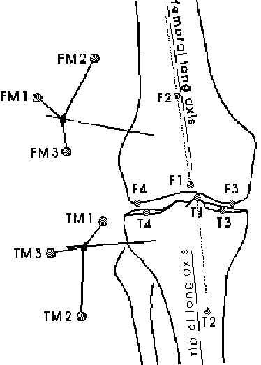
For the tibial coordinate system, the most proximal point of the medial intercondylar eminence (T1, Fig. 21) was selected as the origin of the tibial coordinate system. A second point T2, lying on a line parallel to the longitudinal axis of the tibia and going through T1, was used to define the proximal-distal y-axis. The medio-lateral axis was determined by projecting the estimated centers of the medial (T3) and lateral (T4) tibial articular surfaces onto the plane going through T1 and being perpendicular to the proximal-distal axis. The remaining anterior-posterior axis was then again calculated using the cross product.
Using coordinate transformations (Sderkvist and Wedin, 1993), the femur pin markers were then expressed in the RSA femoral coordinate system, and similarly, the tibia pin markers were expressed in the RSA tibial coordinate system (see also appendix). The following calculations were then exactly as if these coordinates were the coordinates recorded during the standing trial (see chapter 3). The joint translations along the femur fixed flexion axis (medio-lateral shift), along the floating ab/adduction axis (anterior-posterior drawer), and along the tibial fixed internal/external rotation axis (compression/distraction) were calculated. Since the medio-lateral shift and the compression/distraction magnitudes are dependent upon the rotation around the floating axis, these translations were adjusted accordingly (Grood and Suntay, 1983; Lafortune et al. 1992a). The terms medio-lateral shift, and anterior-posterior drawer always refer to translations of the tibia with respect to the femur. The terms medio-lateral shift, anterior-posterior drawer and compression/distraction may not be directly "translated" into the equivalent clinical terms. For instance, compression/distraction does not refer to the compression/distraction experienced between the contact areas of the articulating surfaces of the tibia and femur. Compression/distraction as used in this study refers to the entire tibiofemoral joint structure as being shortened or stretched in a direction parallel to the long axis of the tibia (Lafortune et al., 1992a).
The tibiofemoral flexion/extension curves were fairly similar across the three subjects (Fig. 22, page
*). The knee flexion/extension curves also corresponded well with respect to position at touchdown, amplitude and shape of the curves to another study also assessing skeletal tibiofemoral motion during running (McClay, 1990). Differences across subject (Fig. 22) mainly consisted in the amplitude of the flexion/extension motion and in the position at touchdown. The average position at touchdown for the standing trial based curves (solid lines in Fig. 22) varied from 0 (subject 1) to 15 (subject 3). However, these differences may be partially attributed to alignment uncertainties in defining the axis of the tibial and femoral anatomical coordinate systems. For subject 3, the standing trial and RSA based knee flexion/extension curves agreed very well in amplitude and shape, however, there was a consistent shift between the two curves.The ab/adduction patterns varied considerably between the subjects (Fig. 22). The ab/adduction were similar in subject 3 and 5, whereas the amplitude of this motion was higher for subject 5. Both subjects 3 and 5 showed an initial abduction movement occurring from 0% to 40% of stance followed by an adduction motion until the end of stance. This initial abduction motion averaged 6 for subject 3, and 9 subject 5. On the other hand, subject 1 showed a small initial adduction movement of 4, followed by smaller abduction and adduction motions. Towards the end of stance phase, subject 1 exhibited an average adduction motion in the order of magnitude of 3. As found for flexion/extension, the RSA based and standing trial based ab/adduction agreed well for subject 3, and the only difference consisted again of a constant shift between the two average curves.
The patterns and magnitude of ab/adduction during stance was in total disagreement with the only other running study reporting skeletal knee ab/adduction (McClay, 1990). McClay (1990) found that all four subjects exhibited a similar pattern: an initial adduction motion averaging 6 from touchdown to around 40% of stance, followed by a gradual abduction (8) until the end of stance. None of the subjects in the present study showed a similar pattern. Part of these differences may be attributed to the fact that the coordinate system definitions used in McClay (1990) were based on RSA whereas in this study they were based on a neutral standing trial. However, interestingly even the RSA based curves for subject 3 did not agree at all with McClay’s data, although the anatomical reference frames for both the femur and tibia were defined using exactly the same method.
Due to the ligamentous restrictions imposed by the medial and lateral collateral ligaments and due to the geometry of the knee joint, the ab/adduction motion at the knee joint is very limited. The range of motion of knee ab/adduction is approximately 5, whereas the possible range of motion of the other rotations are much higher; the ranges of motion of flexion/extension and internal/external knee rotation are approximately 150 and 35, respectively (Frank et al., 1995). It can be speculated that the range of motion of ab/adduction is even smaller during running when the knee joint is loaded and stabilized by muscular forces. However, the ranges of motion in ab/adduction for some subjects found in this study and in the study by McClay (1990) well exceeded 5. This suggests that the ab/adduction motion measured may not be the "true" ab/adduction motion. Due to the small magnitude, the ab/adduction motions are highly influenced by alignment problems of the anatomical coordinate systems. The effect of alignment problems and resulting cross-talk may exceed and mask the actual ab/adduction motion. In this context, it is interesting to note that in the two subjects showing a considerable amount of ab/adduction (subjects 3 & 5, Fig. 22), the patterns of ab/adduction were very similar to the flexion/extension motion giving rise to speculations that the relatively large ab/adduction motions were mainly caused by cross-talk.
The internal/external knee rotation patterns were fairly similar across subjects. From touchdown to midstance, all subjects showed an initial internal knee rotation (Fig. 22). However, the magnitude of that motion ranged considerably between subjects: Almost no initial knee rotation was present in subject 5 (2), whereas the initial internal knee rotation was very pronounced in the other two subjects (9 for subject 1), 7 for subject 3). The subjects exhibited an external knee rotation during the second half of stance. For all subjects, this external knee rotation towards the end of stance exceeded the initial internal knee rotation leaving the knee in a more externally rotated position at take-off as compared to touchdown. The patterns and magnitudes of internal/external knee rotation found in this study compared favourably to the patterns and magnitudes presented in the study by McClay (1990) with the exception that in that study the magnitude of the initial internal knee rotations was higher than the magnitude of external knee rotation during the second half of stance.
Again, it has to be realized that at least part of the differences found between subjects for the internal/external knee rotations may be attributed to small deviations and inconsistencies in defining the anatomical coordinate systems and resulting cross-talk. For subject 3, the internal/external knee rotation curves based on RSA and the neutral standing trial were again very similar and an obvious shift between the two average curves can be reported (Fig. 22). However, in contrast to flexion/extension and ab/adduction, there was a noticeable difference in amplitude; the initial internal knee rotation based on RSA and the neutral standing trial averaged 11 and 7, respectively.
On a different note, one of the five trials in subject 5 appeared to be an outlier (Fig. 22). Since this particular trial was trial number 1 in the series of the five trials, it was first suspected that between trial 1 and the following four trials the femur pin may have moved. However, the same trial also produced an "outlier" for the skin marker based curves (see Fig. 15), and therefore, the possibility of a moving (femur) pin between these trials was excluded.
When comparing the ab/adduction and internal/external knee rotation curves with the flexion/extension curves within the same subject, it is apparent that most of these curves resemble the flexion/extension patterns. In other words, the ab/adduction and internal/external knee rotation curves are the same as the flexion/extension curves with different amplitudes. It appeared that only the ab/adduction in subject 1 and the internal/external knee rotation in subject 5 did not have this "pattern agreement". The similarities between the flexion/extension curves and the other curves within the same subject suggested that there may have been some cross-talk. On the other hand, it may also be argued that the knee rotations are coupled, in the sense that for instance, an internal knee rotation takes place when the knee undergoes a flexion motion (Blankevoort et al., 1988). Based on the data presented in this study, it is difficult to estimate how much of the non-primary knee rotations (ab/adduction, internal/external knee rotation) is due to a "real" rotation and how much can be attributed to alignment problems of the anatomical reference frames. More research is needed to establish reliable and meaningful coordinate systems in order to make valid comparisons across subjects. On the other hand, it may also be argued that Cardan angles using a joint coordinate system (Grood and Suntay, 1983) may not be appropriate to determine knee rotations other than flexion/extension.
The tibiofemoral joint translations of the five trials for subject 3 are depicted in Fig. 23. Since the origin of the femoral and tibial anatomical coordinate systems did not have coincident origins, there is an "off-set" present in these graphs. In other words, already in a "neutral" position the origins of the two reference frames would be some distance apart from each other. Therefore, the change in movement should be considered rather than the absolute values displayed on the vertical axes (Fig. 23).
The anterior/posterior drawer curves indicate that during the first 5% of stance almost no movement took place (Fig. 23). Thereafter, the origin of the tibial reference frame moved 4 mm posteriorly with respect to the femur. From around 40% to 80% of stance, an anterior movement of the tibia (5 mm) took place, followed by a fast posterior movement of the tibia towards the end of stance.
The distraction/compression pattern was very similar to the anterior/posterior drawer pattern (Fig. 23). Almost no compression/distraction movement took place during the 10% of stance. From 10% to 40% of stance, a distraction movement (5.6 mm) took place between the origins of the tibial and femoral reference frame. This distraction movement was followed by a larger compression movement (6.8 mm). During the final 20% of stance, a distraction movement of 2.8 mm was observed between the origins of the two coordinate systems.
The medio-lateral shift was the translatory motion with the least amount of movement excursion during stance (Fig. 23). No consistent medio-lateral motions across the five single trials was present during the first 15% of stance. Thereafter, the tibia underwent a lateral shift averaging 3.6 mm. From 40% to 80% of stance, the origin of the tibial reference frame shifted medially (4.1 mm) with respect to the femoral reference frame. During the last 20% of stance, the tibia shifted again in a lateral direction (1.8 mm).
The patterns found for anterior-posterior drawer and distraction/compression for subject 3 were similar to the patterns reported by McClay (1990). The pattern of medio-lateral shifting was however not in agreement with the data presented by McClay (1990). The patterns of medio-lateral shifting in this study were generally in the opposite direction to what has been reported by McClay (1990). This discrepancy may be related to the discrepancies found for the ab/adduction motion (see page
*).All tibiofemoral translations (Fig. 23) exhibited a striking similarity with the knee flexion extension behaviour (Fig. 22). This has also been found in previous investigations determining the skeletal tibiofemoral translations during walking and running (Lafortune, 1984; McClay, 1990). The striking similarity between the translations and the knee flexion is not surprising. Even though the orientation and location of the anatomical tibial and femoral reference frames were based on anatomical considerations, the points chosen as the origins of the femoral and tibial reference frames can be considered to be "arbitrary". That means that they are likely not to reflect an "average" knee joint center. If the origins of the two coordinate systems do not coincide with an "average" joint center or if such an "average" joint center does not exist, the translations are very much dependent on the rotations. In other words, cross-talk exists in the sense that translations would be registered even though a pure rotation took place. The methodological concerns regarding the sensitivity of the tibiofemoral translations with respect to the definition of the anatomical coordinate systems has been pointed out previously (Blankevoort et al., 1988). Blankevoort and co-workers (1988) suggested that relative translations of a point on the femur with respect to a point located at the tibia would be more meaningful than translations calculated along the axis of a joint coordinate system.
The joint translations as calculated for subject 3 (Fig. 23) will not be further discussed since it is believed that these results are primarily a function on how the anatomical coordinate systems were defined, and the physiological meaning of these results are limited. For a comprehensive interpretation of the joint translations, meaningful distances between points located in the femur and tibia should be calculated (Blankevoort et al, 1988). This was attempted in the present study by calculating the distance between average femoral and tibial attachment sites of the anterior cruciate ligament (ACL). These points were identified and digitized on the radiographic pictures (RSA) which allowed the calculation of the distance between these points during the stance phase of running. As expected, the translations were much smaller than the translations shown in Fig. 23. However, the changes in distance between the average femoral and tibial ACL attachment sites were in the range of the accuracy of the motion measurement system used (1.5 mm), and therefore, the author did not feel comfortable presenting that data. In order to calculate such small changes, a closer camera view of the knee joint would have been required to attain a better accuracy enabling to calculate such small changes.
The knee rotations were presented for three subjects and the knee translations for one subject during the stance phase of running. It was found that the non-primary knee rotations (ab/adduction, internal/external knee rotation) as well as the translations were highly influenced by the knee flexion/extension patterns during the stance phase of running. Based on the results of this study, it cannot be concluded how much of these rotations and translations were "real" and how much was due to the definition of the anatomical coordinate systems used in this investigation ("cross-talk"). Further research is needed to establish reliable, reproducible and meaningful anatomical coordinate systems for both femur and tibia. It can also be argued that Cardan angles calculated using joint coordinate systems (JCS) may not be the method of choice to determine the non-primary knee rotations. It was concluded that joint translations determined using a JCS may only have limited physiological meaning. For future studies, it is suggested to calculate meaningful distances between points located in the two bodies, such as the insertion sites of ligaments. Such distances are believed to provide a more comprehensive and physiologically meaningful translations than translations measured using a joint coordinate system.
CHAPTER 7:
Implications for Future Studies Using External Markers
The purpose of this chapter is (a) to discuss the implications of the results of this project with respect to future studies using external markers to estimate the skeletal joint motions at the lower extremities, and (b) to make suggestions for future research in the area of skin (or external marker) movement artefact. The implications and possible future work will be discussed separately for the shoe/foot (calcaneus), the shank (tibia) and the thigh (femur).
The results of the previous chapters (chapters 3&4) have shown that:
- External markers indicate the tibiocalcaneal rotations, but overestimate the skeletal kinematics.
- A relationship appears to exist between variables extracted from external and skeletal markers (
These findings imply that shoe markers may not provide an accurate representation of the skeletal calcaneal kinematics. Hence, skeletal kinematics should not be predicted from markers attached to the shoe. Based on the results of this study and based on the review of literature, two approaches (considerations) are suggested for future studies:
- The differences between the variables determined from bone and shoe markers (Fig. 11, page
As long as this has not been shown, one may assume that increased bone movement does results in increased shoe movement and that trends may be quantified. Inter-shoe comparisons, however, must be considered with caution. Small kinematic differences between different shoes may either reflect true skeletal differences or such differences may just be a reflection of more or less relative movement (slipping) occurring between the calcaneus and the shoe.
The results of this project have shown that for both the tibiocalcaneal (ankle-joint complex) and tibiofemoral (knee) rotations, the skin movement artefact caused by the shank markers was almost negligible in comparison to the external marker movement artefact caused by the shoe or the thigh markers. In order to find out how accurately the rotations of the tibia can actually be determined with skin markers, the tibial rotations were calculated with respect to the laboratory coordinate system both based on external and skeletal markers.
Tibial rotations during running were calculated for the five subjects for which AJC (tibiocalcaneal) rotations were presented in chapter 4. The rotations of the tibia with respect to the laboratory coordinate system were determined using Cardan angles. For these calculations, the longitudinal tibial rotation occurred around the proximal-distal (longitudinal) axis of the tibia, tibial ab/adduction around the floating axis, and tibial flexion/extension around the z-axis (axis perpendicular to running direction and vertical laboratory axis) of the laboratory (or film) coordinate system. The kinematics were determined based both on skeletal and skin markers. For the skin markers, all markers were used which were visible throughout the entire stance phase (see chapter 4). That means, that depending on the subject, four shank markers (subject 1), five shank markers (subject 3), or all six shank markers were used (subjects 2, 4 & 5).
In the following pages, only the tibial rotation about the longitudinal axis is presented because the largest differences between skin and bone marker based rotations can be expected to occur about this rotation (Holden et al., 1994), and because this rotation is believed to be important with respect to running injuries (Nigg et al., 1993). For simplicity, the term "tibial rotation" will be used to refer to tibial rotation about the longitudinal axis. Note that the tibial rotation as used in the following is the rotation of the tibia with respect to the laboratory coordinate system, and is not synonymous to internal /external tibial rotation or leg rotation as discussed in the literature review of this thesis (see page
*). "Internal/external tibial (or leg) rotation" as presented in the literature review refers to the rotation of the tibia (leg) with respect to the foot.During midstance, when the foot is on the ground, the rotation of tibia with respect to the laboratory coordinate system and with respect to the foot can be expected to be very similar.
Longitudinal Tibial Rotation |
Sub. 1 |
Sub. 2 |
Sub. 3 |
Sub. 4 |
Sub. 5 |
Mean |
RMS Diff. [ ]Max. Diff. [ ]RMS Diff. [%] Max. Diff. [%] |
0.8 2.5 5.0 16.1 |
1.8 2.8 14.6 22.4 |
1.6 2.7 13.2 22.1 |
1.5 3.2 14.2 29.5 |
0.9 1.6 7.5 13.9 |
1.3 2.6 10.9 20.8 |
The longitudinal tibial rotation based on skeletal and bone markers are depicted in Fig. 24. For all subjects, excellent agreement between the skin and bone marker based tibial rotation was found with respect to the amplitude as well as with respect to the shape of the curves (Fig. 24). This was also reflected in the small mean (RMS) differences and maximal differences between the skeletal and skin marker derived tibial rotations (Table 9).
The differences found in this study between skin and bone marker based tibial rotations were slightly lower than the differences found in a previous study comparing skin and bone marker based tibial rotations during walking (Holden et al., 1994). From the curves of the three subject presented in Holden et al. (1994), the maximal difference during stance was estimated to be 3.8 (averaged across the three subjects), whereas in the present study the maximal differences averaged 2.6 (Table 9). It is surprising that the differences were actually lower for running (present study) than for walking (Holden et al., 1994). However, it is suggested that the smaller difference for running can be explained by subject differences and differences in number and placement of skin markers used rather than by the different movements (running vs. walking).
The results of this study have shown that rotation of the tibia along its longitudinal axis is well represented with the use of skin markers as employed in this study. It is speculated that the representation of the skeletal tibial rotation could even be improved when all skin markers would be placed directly over the tibia instead of also placing markers over other structures, i.e. the fibula (lateral shank markers: S1 to S3, see Fig. 1).
The results of this project have shown that skin movement artefact occurring at the thigh is a major problem. Rotations other than flexion/extension cannot be reliably determined when using skin markers.
For the results presented in the previous chapters, five skin markers (Th2 to Th6) were used to calculate the skin marker based rotations. However, some markers may move more than others with respect to the underlying bone, and therefore, a better representation of the skeletal kinematics may be obtained when excluding such ("bad") markers from the calculations. The purpose of the following paragraphs is to come up with some recommendations regarding marker placements at the thigh. For that purpose, two type of analyses were performed, a quantitative analysis and a qualitative analysis.
Quantitative Analysis of Movement Artefact of Thigh Markers
The data for the running trials of the three subjects for which valid femur pin data was available were used for this analysis (same data as used for chapter 5). For these trials, all possible marker combinations (3 number of markers 5) of the five thigh markers (Th2 to Th6) were used to determine which marker combination delivered the best representation of the skeletal kinematics. The quality of a certain marker combination was quantified by using a similar analysis as the segmental error analysis (see page
*). For each trial, the difference between the tibia-femur and the tibia-thigh rotations were calculated during the entire stance phase indicating how well the tibiofemoral kinematics were represented with the specific thigh marker combination. The segmental thigh error was than averaged for all rotations, trials and subjects. This value was termed mean thigh error (MTE).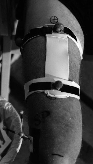
In addition to the different thigh marker combination, the MTE was also calculated for three running trials with shoe 4 (see Fig. 17) for the three subjects with valid femur pin data. For these running trials, the subjects wore a thigh skin frame (Fig. 25). The center part of the frame (arranged in a proximal-distal direction) consisted of a strip of plastic (Sanssplint™) which was 20 cm in length and 5 cm in width. At the each end of this strip, separate strips of thermoplast were secured, encompassing most of the thigh. Three skin markers were attached to the frame as depicted in Fig. 25. The skin frame was attached to the thigh with double sided tape and additionally, the distal and proximal "circumferential" strips were closed at the posterior part with tape. Prior to the motion recordings and the insertion of the bone pins, the thigh frame was fitted to each of the subjects in order to ensure an optimal fit.
A second analysis was also performed to assess how much each thigh marker moved with respect to the underlying femur (using again the same data as used for chapter 5). For this analysis, all of the thigh marker positions were expressed in a femur coordinate system based on the three markers attached to the pins. The difference of these thigh marker positions during the stance phase compared to during the standing trials was determined as a measure on how much these markers moved at each instant in time during the stance of the running trials. An average relative marker movement (RMM) of each marker was then calculated during the stance phase from all the five running trials and from all the three subjects (subjects 1, 3, & 5) for which valid femur pin data was available.
Marker Combination |
MTE |
Rank |
||||
Th2 |
Th3 |
Th4 |
Th5 |
Th6 |
[] |
|
4 |
4 |
4 |
4 |
4 |
3.7 |
8 |
4 |
4 |
4 |
4 |
3.9 |
10 |
|
4 |
4 |
4 |
4 |
3.8 |
9 |
|
4 |
4 |
4 |
4 |
3.1 |
5 |
|
4 |
4 |
4 |
4 |
4.4 |
13 |
|
4 |
4 |
4 |
4 |
2.9 |
2 |
|
4 |
4 |
4 |
4.6 |
15 |
||
4 |
4 |
4 |
3.3 |
6 |
||
4 |
4 |
4 |
2.7 |
1 |
||
4 |
4 |
4 |
4.9 |
16 |
||
4 |
4 |
4 |
4.5 |
14 |
||
4 |
4 |
4 |
4.1 |
12 |
||
4 |
4 |
4 |
3.3 |
7 |
||
4 |
4 |
4 |
3.0 |
4 |
||
4 |
4 |
4 |
2.9 |
3 |
||
4 |
4 |
4 |
11.1 |
17 |
||
Skin Frame |
4.0 |
11 |
||||
The results of the mean thigh errors in terms of rotations are presented in Table 10. All MTE values were between 2 and 5 except when using the thigh marker combination Th4-Th5-Th6. The results for the Th4-Th5-Th6 combination were excepted as these markers were more or less on a line (colinear), which results in a poor description of a rigid body in space. The best skin marker combination was Th2-Th3-Th6. However, this combination was only marginally better than other combinations. The skin frame used did not produce less error. However, it must be mentioned that this skin frame was used during different running trials with a different shoe (shoe 4). Shoe 4 had a much stiffer midsole than the shoe 1. This may have affected the amount of the skin movement artefact at the thigh and therefore these results cannot be compared directly.
Marker |
RMM [mm] |
Th1 |
32.8* |
Th2 |
14.5 |
Th3 |
7.7 |
Th4 |
26.5* |
Th5 |
16.9 |
Th6 |
15.3 |
The results of the relative movement of each of the thigh markers with respect to the underlying bone confirmed the results found for the rotational errors of the different possible combinations as presented in Table 10. Markers Th2, Th3 and Th6 had also the least amount of RMM (Table 11). This error analysis is limited as errors can be expected to increase the further away the markers are from the location of the bone markers used to define the skeletal femoral coordinate system. This increase in error is due to two reasons. First, small inaccuracies (caused e.g. by digitizing errors) in the definition of the directions of the bone-embedded femoral coordinate system are amplified the further away the markers are. For instance, a 2 rotational error in defining the femoral coordinate system causes an error of 1.7 mm (= sin(2) * 50 mm) for a marker at a distance of 5 cm. On the other hand, if the marker is 40 cm away, such a rotational deviation would result in 14 mm error in determining the RMM of such a marker. Secondly, the further the thigh markers are away from the bone markers, the more they are affected by the inaccuracies in the DLT calculations (outside calibration volume). Due to these reasons, it is suggested that the results (RMM) of the markers furthest away from the bone markers are not reliable (Th1, Th3).
One problem inherent with both quantitative variables used (MTE and RMM), is that results may be masked or affected by the DLT inaccuracies of the thigh markers. Due to this limitation, recommendations with respect to marker placements at the thigh could not be made exclusively based on these analyses. Hence, in addition to the quantitative analysis, a qualitative analysis was performed.
Qualitative Analysis of Movement Artefact of Thigh Markers
For the qualitative analysis, all the running trials used in this study were examined carefully on the films. The following observations were made:
- Generally, the lateral markers (Th1, Th2, Th3) appeared to be more stable than the markers placed in front of the thigh (Th4, Th5, Th6). This observation was in agreement with the finding of Cappozzo et al. (1996).
- The thigh marker Th4 (middle front marker) showed generally the greatest wobbling of all thigh markers.
- The marker placed at the greater trochanter appeared to be the best marker based on the observation of the films. This finding is in disagreement with the RMM results (Table 11), however as mentioned earlier, it is very likely that the RMM results for this marker cannot be trusted (DLT problems, large distance from bone markers). Interestingly, the observation that the greater trochanter marker appeared to be a good location is somewhat in disagreement to the conclusions made in a previous study (Cappozzo et al., 1996). Cappozzo and co-workers (1996) suggested that skin markers above anatomical landmarks (including the greater trochanter) are unsuitable for marker placements.
- Based on the observation of the film, the following ranking was made (from best to worst):
- Th1
- Th3 and Th6
- Th2 and Th4
- Th5
- Interestingly, the marker placed in the middle front (Th5) showing the most relative movement (wobbling) with respect to the underlying also appeared to influence the adjacent marker on the lateral side (Th2).
- The skin frame reduced the relative movement between the markers attached to it. However, the skin frame appeared to move as one unit with respect to the underlying bone, especially during the period when the thigh muscles were activated.
- The key reason for the above observations appeared to be the location of muscles and muscle activities. Markers placed on tendinous structures (Th3, Th6) appeared to be more stable than markers placed directly on muscles (Th5 placed on the muscle belly of the rectus femoris). Based on these considerations, locations on muscles (muscle bellies) should be avoided particularly if the muscles are rather large and are activated and deactivated during the movement in consideration.
The use of skin markers at the thigh appears to be very problematic when accurate three-dimensional kinematics is required. The limited results as presented in the previous paragraphs suggest that markers should not be placed on muscles that are activated during the movement in consideration. It is suggested that for any type of kinematic analysis involving the thigh, a number of markers (grid of markers) should be attached to the thigh and the subject(s) should be filmed or videotaped during the movement. A simple qualitative analysis (as performed in this study) may help to exclude obviously poor marker placements which introduce large differences between external and skeletal kinematics.
As for running, it is suggested that the markers Th2-Th3-Th6 provide the best (although still a rather poor) representation of the skeletal femoral kinematics. Markers placed directly over the muscle belly of the rectus femoris (Th5) should definitely be avoided.
Conclusions and Future Research
Further research is needed to gain a better understanding of the skin movement artefact at the thigh and to establish methods that are able to provide an improved representation of skeletal (femoral) kinematics during highly dynamic movements such as running. For that purpose, future studies should be conducted which focus on the skin movement artefact at the thigh, and evaluate a greater number of skin markers to create recommendations for suitable skin marker locations on the thigh.
The author questions whether external markers, even when used in conjunction with a correction algorithm or a method to reduce the skin movement artefact, can (ever) be used to obtain an accurate representation of the skeletal femoral kinematics during a highly dynamic movement such as running. In that sense, the development of minimally invasive in-vivo measurements as for instance suggested by Tashman and co-workers (1996) may be a more promising avenue for future research aimed at accurate kinematic assessments involving the femur.
CHAPTER 8: Summary and Conclusions
The objectives of this study were to determine the three-dimensional skeletal tibiocalcaneal (ankle joint complex, AJC) and tibiofemoral (knee) motion during the stance phase of walking and running with shoes, and to compare this to the respective motion determined from external markers. Five male subjects (age 28.6 4.3 years) participated in the study. All subjects were injury free at the time of testing and none of the subjects had an injury history which may have resulted in an abnormal gait. Under local anesthesia, intracortical bone pins (2.5 mm diameter) were inserted into the lateral femoral and tibial condyles, and into the postero-lateral aspect of the calcaneus of the subject’s right leg. Triads of reflective markers were attached to these pins, and additionally, six skin markers were attached to the thigh, six skin markers to the shank, and three markers were fixed to the shoe. For each subject, three walking and five running trials were recorded during the stance phase of walking and running using three high speed cine cameras (LOCAM) operating at 50 Hz for walking and 200 Hz for running. For these trials, the subjects wore standard running shoes (Adidas Equipment Cushioning 1994) without socks. The position of the simultaneously collected skeletal and externally mounted markers were digitized manually for each of the cameras. The three-dimensional coordinates of all the markers were calculated using a standard DLT (direct linear transformation) approach. A "neutral" standing trial collected prior to the motion trials was used to define the anatomical references frames of the various segments. Additionally, a set of radiographs were also taken either prior or after the motion measurements enabling the additional definition of RSA (roentgen-stereophotogrammetric analysis) based anatomical coordinate systems for the femur and tibia. Cardan angles (and respective translations) were used to express the intersegmental knee and AJC motion. Knee flexion/extension, ab/adduction, and internal/external knee rotation as well as AJC plantar/dorsiflexion, ab/adduction and in/eversion were calculated both from skeletal and external markers.
The comparison between skin marker based kinematics of bone pin trials and "dry runs" collected prior to the insertion of the pins revealed that the running style of the subjects was not significantly affected by the insertion of the bone pins. Problems with the femur pin attachment were encountered in some subjects. For one subject, a loosening of the pin was detected during the experiment whereas for another subject a non-rigid femur pin attachment was detected when observing the films prior to data analysis. Consequently, tibiofemoral motion based on skin and skeletal markers were only determined from the remaining three subjects. An analysis of all the standing trials collected during the course of the experiment also suggested that the femur pin drifted (rotational drift) in two of the remaining subjects. However, this rotational drift was small for the time span during which data for this project was collected. Therefore, these two subjects could still be included in the analysis.
For walking and running, the skeletal tibiofemoral (AJC) rotations were generally well reflected with external markers. However, the skeletal rotations were typically overestimated when using external markers. For instance, during running, the maximal initial eversion occurring from touchdown to midstance averaged 16.0 when using external markers. However, the same variable determined from skeletal markers was only 8.6. A segmental error analysis revealed that the differences found between the external and bone marker based kinematics were primarily caused by the relative movement of the shoe markers with respect to the underlying calcaneus. It was speculated that the artefact caused by the relative movement between shoe and calcaneus was mainly caused by the relative movement between shoe and heel and not between heel and calcaneus. Therefore, heel markers tracked through windows cut into the shoe may provide a better estimate of the calcaneal motion than markers directly attached to the shoe as used in this study.
Both during walking and running, good agreement was found between knee flexion/extension derived from skin and bone markers. For the other knee rotations however, the agreement between the rotations calculated from external and skeletal markers ranged from good to virtually no agreement. For ab/adduction and internal/external knee rotations, the differences were large considering the small amplitude of these rotations, and in some subjects, these errors exceeded the actual skeletal motion. A segmental error analysis showed that the discrepancies between external and skeletal kinematics was mainly caused by the skin movement artefact at the thigh. The errors caused by the skin movement artefact at the shank were almost negligible in comparison to the skin movement artefact at the thigh.
Methodological problems were also identified concerning the use of Cardan angles to describe the three-dimensional motion of the tibiofemoral joint. Internal/external knee rotation and particularly ab/adduction can be expected to be small, and these rotations are highly influenced by cross-talk (from knee flexion/extension) resulting from alignment problems of the anatomical coordinate systems. For instance, 5.6 ab/adduction and 6.7 internal/external knee rotation would be registered during a pure knee flexion motion of 30 if the uncertainty in the defining the anatomical coordinate system (of either the femur or tibia) was only 6 for internal/external knee rotation and 3 for ab/adduction. The values used for this specific example correspond to estimated values for knee flexion occurring during the stance phase of running (30 flexion), and to uncertainties in alignment (6 in internal/external knee rotation, 3 for ab/adduction) when using the procedures employed in this project.
Problems were also identified with the representation of joint translations using a joint coordinate system. These translations highly depend on the definition of the anatomical coordinate systems. Additionally, these translations are also highly dependent on the knee flexion/extension motion. Therefore, the physiological meaning of such joint translations may be questioned. For future studies, it is suggested to calculate meaningful distances between points embedded in both segments such as distances between femoral and tibial insertion sites of ligaments.
Based on the results of this project, it was concluded that during walking and running:
- tibiocalcaneal rotations are generally well represented with the use of external markers, but absolute values have to be interpreted with caution since the tibiocalcaneal rotations can be expected to be overestimated when using external markers, and that
- knee rotations other than flexion/extension may be affected with substantial errors when using skin markers.
Abdel-Aziz, Y.I., and Karara, H.M. (1971) Direct linear transformation from comparator coordinates into object space coordinates in close-range photogrammetry. In ASP Symp. on Close Range Photogrammetry. American Society of Photogrammetry, Falls Church, pp. 1-19.
An, K.-N., Jacobsen. L.J., Berglund, L.J., and Chao, E.Y.S. (1988) Application of a magnetic tracking device to kinesiologic studies. J. Biomechanics 21, 613-620.
Andriacchi, T.P., and Toney, M.K. (1995) In-vivo measurement of six-degrees-of-freedom knee movement during functional testing. Transactions of the 41st Annual Meeting of the Orthopaedic Research Society, Orlando, Florida, p. 698.
Angeloni, C., Cappozzo, A., Catani, F., and Leardini, A. (1993) Quantification of relative displacement of skin- and plate-mounted markers with respect to bones. J. Biomechanics 26, 864.
Areblad, M., Nigg, B.M., Ekstrand, J., Olsson, K.O., and Ekstrm, H. (1990) Three-dimensional measurement of rearfoot motion during running. J. Biomechanics 23, 933-940.
Blankevoort, L., Huiskes, R., and Lange, A. de (1988) The envelope of passive knee joint motion. J. Biomechanics 21, 705-720.
Blankevoort, L., Huiskes, R., and Lange, A. de (1990) Helical axes of passive knee joint motions. J. Biomechanics 23, 1219-1229.
Bogert, A.J. van den, Weeren, P.R. van, and Schamhardt, H.C. (1990) Correction for skin displacement errors in movement analysis of the horse. J. Biomechanics 23, 97-101.
Biomch-l (1995) Various discussions regarding recommendations for standardization in the reporting of kinematic data. Biomch-l is an e-mail discussion forum. Archives can be searched at http://www.kin.ucalgary.ca/isb/biomch-l.html.
Cappozzo, A., Catani, F., Della Croce, U., and Leardini, A. (1995) Position and orientation in space of bones during movement: anatomical frame definition and determination. Clin Biomech 10, 171-178.
Cappozzo, A, Catani, F., and Leardini, A., Benedetti, M.G., and Della Croce, U. (1996) Position and orientation in space of bones during movement: experimental artefacts. Clin Biomech 11, 90-100.
Chze, L., Fregly, B.J., and Dimnet, J. (1995) A solidification procedure to facilitate kinematic analysis based on video motion. J. Biomechanics 28, 879-884.
Clement, D.B., Taunton, J.E., and, Smart, G.W. (1984) Achilles tendinitis and peritendinitis: Etiology and treatment. Am. J. Sports Med. 12, 179-184.
Cole, G.K., Nigg, B.M., Ronsky, J.L., and Yeadon, M.R. (1993) Application of the joint coordinate system to three-dimensional joint attitude and movement representation: a standardization proposal. J. biomech. Engng 115, 344-349.
Delozier, G.S., Alexander, I.J., and Narayanaswamy, R. (1991) A method for measurement of integrated foot kinematics. In Proceedings of the International Symposium on 3-D Analysis of Human Movement. Montreal, Canada, pp. 52-54.
Edington, J., Frederick, E.C., and Hamill, C. (1990) The study of rearfoot movement in running. In Biomechanics of distance running (Edited by Cavanagh, P.R.), pp. 135-164. Human Kinetics, Champaign, IL.
Engsberg, J.R., and Andrews, J.G. (1987) Kinematic analysis of the talocalcaneal/ talocrural joint during running support. Med. Sci. Sports 19, 275-284.
Frank, C.B., and Shrive, N.G. (1995) Biomaterials: Ligaments. In Biomechanics of the musculo-skeletal system (Edited by Nigg, B.M., and Herzog, W.), pp. 106-132. John Wiley & Sons, New York.
Gheluwe, B. Van, Tielemans, R., and Roosen, P. (1995) The influence of heel counter rigidity on rearfoot motion during running. J. Appl. Biomech. 11, 47-67.
Grood, E.W., and Suntay, W.J. (1983) A joint coordinate system for the clinical description of three-dimensional motions: applications to the knee. J. biomech. Engng 105, 136-144.
Hatze, H. (1988) High-precision three-dimensional photogrammetric calibration and object space reconstruction using a modified DLT-approach. J. Biomechanics 21, 533-538.
Holden, J.P., Orsini, J.A., and Stanhope, S.J. (1994a) Estimates of skeletal motion: movement of surface-mounted targets relative to bone during gait. In Proceedings of the Thirteenth Southern Biomedical Engineering Conference. University of the District of Columbia, Washington, DC.
Holden, J.P., Orsini, J.A., and Stanhope, S.J. (1994a) Skeletal motion estimates. Effect of surface target techniques. In Proceedings of the 2nd World Congress of Biomechanics, Amsterdam, The Netherlands, p. 149.
Inman, V.T. (1976) The joints of the ankle. Williams and Wilkins, Baltimore.
James, S.L., Bates, B.T., and Osternig L.R. (1978) Injuries to runners. Am J. Sports Med 6, 40-50.
James, S.L., and Jones, D.C. (1990) Biomechanical aspects of distance running injuries. In Biomechanics of distance running (Edited by Cavanagh, P.R.), pp. 249-270. Human Kinetics, Champaign, IL.
Kadaba, M.P., Ramakrishnan, H.K., and Wootten, M.E. (1990) Measurement of lower extremity kinematics during level walking. J. Orthop. Res. 8, 383-392.
Karlsson, D., and Lundberg, A. (1994) Accuracy estimation of kinematic data derived from bone anchored external markers. In Proceedings of the 3rd International Symposium on 3-D Analysis of Human Motion. Hasselbacken Conference Center Stockholm, Sweden, pp. 27-30.
Karlsson, J.O.M. (1990) Using axiodes to compare biokinematic data measured with bone- and skin-mounted markers. MA Thesis, Massachusetts Institute of Technology, Boston.
Kennedy, P.W., Wright, D.L., and Smith, G.A. (1989) Comparison of film and video techniques for three-dimensional DLT repredictions. Int. J. Sports Biomech. 5, 457-460.
Kepple, T.M., Stanhope, S.J., Lohmann, K.N., and Roman, N.L. (1990) A video-based technique for measuring ankle-subtalar motion during stance. J Biomed Eng 12, 253-260.
Koh, T.J., Grabiner, M.D., and de Swart, R.J. (1992) In vivo tracking of the human patella. J. Biomechanics 25, 637-643.
Lafortune, M.A. (1984) The use of intra-cortical pins to measure the motion of the knee joint during walking. Unpublished doctoral thesis, The Pennsylvania State University.
Lafortune, M.A., Cavanagh, P.R., Sommer, H.J. III and Kalenak, A. (1992a) Three-dimensional kinematics of the human knee during walking. J. Biomechanics 25, 347-357.
Lafortune, M.A., Lambert, C., and Lake, M. (1992b) Skin marker displacement at the knee joint. In Proceedings of the Second North American Congress on Biomechanics, Chicago.
Lafortune, M.A., Cavanagh, P.R., Sommer, H.J. III and Kalenak, A. (1994) Foot inversion-eversion and knee kinematics during walking. J. Orthop. Res. 12, 412-420.
Lenox, J.B., and Cuzzi, J.R. (1978) Accurately characterizing a measured change in configuration. ASME Paper No. 78-DETY-50.
Levens, A.S., Inman, V.T., and Blosser, J.A. (1948) Transverse rotation of the segments of the lower extremity in locomotion. J. Bone Jt Surg 30A, 859-872.
Lundberg, A. (1989) Kinematics of the ankle and foot: in vivo roentgen stereophotogrammetry. Acta Orthop. Scand. 60, Suppl. 233, 1-26.
Maslen, B.A., and Ackland, T.R. (1994) Radiographic study of skin displacement errors in the foot and ankle during standing. Clin. Biomech. 9, 291-296.
McClay, I.S. (1990) A comparison of tibiofemoral and patellofemoral joint motion in runners with and without patellofemoral pain. Unpublished doctoral thesis, The Pennsylvania State University.
McClay, I.S., and Manal, K. (1995) Lower extremity kinematic comparisons between forefoot and rearfoot strikers. In Proceedings of the 19th annual conference of the American Society of Biomechanics. Stanford, California. pp. 211-212.
Messier, S.P., and Pittala, K.A. (1988) Etiologic factors associated with selected running injuries. Med. Sci. Sports 20, 501-505.
Moseley, L., Smith, R., Hunt, A., and Gant, R. (1996) Three-dimensional kinematics of the rearfoot during the stance phase of walking in normal young adult males. Clin Biomech 11, 39-45.
Murphy, M.C. (1990) Geometry and the kinematics of the normal human knee. Unpublished doctoral thesis, Massachusetts Institute of Technology, Boston.
Nigg, B.M. (1986) Experimental techniques used in running shoe research. In Biomechanics of running shoes (Edited by Nigg, B.M.), pp. 27-62. Human Kinetics Publishers, Champaign, IL.
Nigg, B.M., and Morlock, M.M. (1987) The influence of lateral heel flare of running shoes on pronation and impact forces. Med. Sci. Sports 19, 294-302.
Nigg, B.M., and Cole, G.K. (1995) Measuring techniques: optical methods. In Biomechanics of the musculo-skeletal system (Edited by Nigg, B.M., and Herzog, W.), pp. 254-286. John Wiley & Sons, New York.
Nigg, B.M., Cole, G.K., and Nachbauer, W. (1993) Effects of arch height of the foot on angular motion of the lower extremities in running. J. Biomechanics 26, 909-916.
Pennock, G.R., and Clark, K.J. (1990) An anatomy-based coordinate system for the description of kinematic displacements in the human knee. J. Biomechanics 23, 1209-1218.
Ramakrishnan, H.K., and Kadaba, M.P. (1991) On the estimation of joint kinematics during gait. J. Biomechanics 10, 969-977.
Reinschmidt, C., Stacoff, A., and Stssi, E. (1992) Heel movement within a court shoe. Med. Sci. Sports 24, 1390-1395.
Ronsky, J.L., and Nigg, B.M. (1991) Error in kinematic data due to marker attachment methods. In Proceedings of XIIIth Congress on Biomechanics. The University of Western Australia, Perth, pp. 390-392.
Shiavi, R., Limbird, T., Frazer, M., Stivers, K., Strauss, A., and Abramovitz, J. (1987) Helical motion analysis of the knee - II. Kinematics of uninjured and injured knees during walking and pivoting. J. Biomechanics 20, 653-665.
Sderkvist, I., and Wedin, P.-. (1993) Determining the movements of the skeleton using well-configured markers. J. Biomechanics 26, 1473-1477.
Soutas-Little, R.W., Beavis, G.C., Vertraete, M.C., and Markus, T.L. (1987) Analysis of foot motion during running using a joint coordinate system. Med. Sci. Sports 19, 285-293.
Stanhope, S.J. (1994) In-vivo measurement of human skeletal motion. In Proceedings of the 2nd World Congress of Biomechanics, Amsterdam, The Netherlands, p. 149.
Spoor, C., and Veldpaus, F. (1980) Rigid body motion calculated from spatial coordinates of markers. J. Biomechanics 21, 45-54.
Stacoff, A., Reinschmidt, C., and Stssi, E. (1992) The movement of the heel within a running shoe. Med. Sci. Sports 24, 695-701.
Tashman, S., DuPr, K., Goitz, H., Lock, T., Kolowich, P., Flynn, M. (1995) A digital radiographic system for determining 3D joint kinematics during movement. In Proceedings of the 19th annual conference of the American Society of Biomechanics. The University of Stanford, Stanford, pp. 249-250.
Taunton, J.E., Clement, D.B., Smart, G.W., Wiley, J.P., and McNicol, K.L. (1985) A triplanar electrogoniometer investigation of running mechanics in runners with compensatory overpronation. Canadian J. Sport Sciences 10, 104-115.
Viitasolo, J.T., and Kvist, M. (1983) Some biomechanical aspects of the foot and ankle in athletes with and without shin splints. Am J. Sports Med 11, 125-130.
Weeren, P.R. van, Jansen, M.O., Bogert, A.J. van den, and Barneveld, A. (1992) A kinematic and strain gauge study of the reciprocal apparatus in the equine hind limb. J. Biomechanics 25, 1291-1301.
Winter, D.A. (1990) Biomechanics and motor control of human movement. John Wiley & Sons, New York.
Wood, G.A., and Marshall, R.N. (1986) The accuracy of DLT extrapolation in three-dimensional film analysis. J. Biomechanics 9, 781-785.
Woltring, H.J., Huiskes, R., Lange, A. de, and Veldpaus, F.E. (1985) Finite centroid and helical axis estimation from noisy landmark measurements in the study of human joint kinematics. J. Biomechanics 18, 379-389.
Woltring, H.J., McClay, I.S., and Cavanagh, P.R. (1989) 3-D camera calibration without a calibration object. In Proceedings of the XII International Society of Biomechanics meeting, Los Angeles, U.S.A.
Woltring, H.J. (1994) 3-D attitude representation of human joints: A standardization proposal. J. Biomechanics 27, 1399-1414.
Wu, G., Cavanagh, P.R. (1995) ISB recommendations for standardization in the reporting of kinematics data. J. Biomechanics 28, 1257-1261.
APPENDIX:
Methods for the Calculation of Rigid Body Kinematics
In the following, the methods which were used to calculate intersegmental joint motions will be described in detail.
X, Y, Z = global coordinates
X = direction of locomotion
Y = vertical
Z = perpendicular to X and Y (right handed coordinate system)
Xi, Yi, Zi = local coordinates in segment i
Xi = anterior-posterior axis (Xi+: anterior)
Yi = proximal-distal axis (Yi+: proximal)
Zi = medio-lateral axis (Zi+: lateral (medial) for the right (left) leg)
Hx, Hy, Hz = relative (translational) orientation between two segments, or in general, between two coordinate systems.
a
, b, g = relative (rotational) orientation between two segments, or more precisely, between two coordinate systems. a, b, g denote rotations around the x, y, and z axes, respectively.r
A = location vector in coordinate system A. rA has the following form:T = transformation matrix:
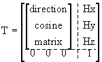
TAB = matrix transforming coordinates from coordinate system "A" to coordinate system "B": rB = [TAB] rA
TAB = sderkvist([rA; rB])†
A = Anatomical coordinate system based on roentgen-stereo analysis (RSA).
S = Anatomical coordinate system based on a neutral position (standing trial).
R = Global coordinate system of the RSA measurements
G = Global (kinematic) coordinate system
ri = rotation around i-axis
Rijk = [rk] * [rj] *[ri] (see also page
*)Segmental coordinate systems (anatomical reference frames) are needed to determine the relative position of two adjacent segments of the human body. For this project, anatomical reference frame were determined with two different methods:
- Neutral Position:
A standing trial or a defined neutral position is used to align the segmental (anatomical) coordinate system to the global (lab) coordinate system. Such coordinate systems cannot be used to calculate meaningful relative translations between two adjacent segments. Furthermore, anatomical coordinate systems that are based on a standing trial do not account for "misalignment" of different segments with respect to each other (e.g. varus/valgus positions).
- Roentgen Stereo Analysis (RSA):
An anatomical reference frame can be determined with the use of digitized landmarks and/or directions in the radiographic reference frame. Anatomical reference frames based on RSA account for subject difference in alignment, but their definition is "arbitrary", i.e. depends on how medio-lateral, proximal-distal, and anterior-posterior axes are defined in the RSA views.
Definition of anatomical coordinate system based on a neutral position
The subject has the segments in a neutral (standing) position where it is assumed that the segments are aligned with the global (laboratory) coordinate system:
rG = rS
Definition of anatomical coordinate system based on RSA
Definition of an anatomical (femur, tibia, and calcaneus) reference frame with the use of digitized anatomical landmarks or directions (in radiographic reference frame).
rA = [TRA] rR
[TRA] can be determined with the use of the "Sderkvist algorithm" (Sderkvist and Wedin, 1993).
Example: sderkvist([0 0 0,1 0 0, 0 1 0; new origin RSA coordinates, point 1/0/0 in new anatom. coord. system expressed in RSA coordinates,...])
 |
The position of the bone markers in each segment can be calculated with the use of [TRA]: r Abone markers = [TRA] rRbone markers |
Determination of absolute and relative orientation of two adjacent segments. As an example, the femur, tibia, thigh and shank segments will be used:
Absolute orientation of a segment
known: rG, rA of the markers
unknown: transformation matrix, TAG
rGsegment = [TAsegmentGsegment] rAsegment
[TAG] ([TAsegmentGsegment]) can again be determined with the use of the "Sderkvist algorithm".
Relative orientation of two adjacent segments
Note that rA stands either for a location vector in (a) an anotomical coordinate system based on a neutral (standing) position or (b) in an anatomical coordinate system based on RSA. For this example, the two adjacent (articulating) segments are the femur and the tibia.
femur (or thigh): r
G = [TAfemurG] rAfemurtibia (or shank): r
G = [TAtibiaG] rAtibiar
Atibia = [TAtibiaG]-1 [TAfemurG] rAfemur[T
AfemurAtibia] rAfemurr
Afemur = [TAfemurG]-1 [TAtibiaG] rAtibia[T
AtibiaAfemur] rAtibiaCALCULATION OF CARDAN ANGLES AND TRANSLATIONS
conventions: X = anterior-posterior axis (Xi+: anterior)
Y = proximal-distal axis (Xi+: proximal)
Z = medio-lateral axis (Zi+: lateral for the right leg, medial for the left leg)
JCS as proposed by Grood and Suntay (1983), and Cole et al. (1993) were used.
Sequence: Z-X-Y (r
(1)
(2)
a: ab/adduction: rotation about the floating axis(3)
b: int./ext. knee rot.: rotation about Y-axis of tibia (tibial-fixed longitudinal axis)Rzxy =
- Determination of
compr./distraction (y) = Hy + sin
medio-lateral shift (z) = Hz + sin
b Hy
The use of the above transformation (femur with respect to (fixed) tibia) leads to the follwing sign conventions:
- flexion/extension:+ flexion
- Ab/Adduction:+ Abduction (- : bow-legged)
- Rotation:+ external tibial rotation with respect to the femur
- a-p drawer:+ posterior drawer of the tibia
- compr./distr.:+ distraction
- m-l shift:+ medial translation of the tibia with respect to the femur
Sequence: Z-Y-X (r
Rzyx =
a, b, and g:Solve for a, b, and g using appropriate elements of the matrix above
Remark: To obtain the skin (external) marker based rotations, "femur", "tibia" and "calcaneus" have just to be replaced with the respective external marker based segments (thigh, shank, shoe/foot).
Note that:
ri = rotation around i-axis
a
,b,g = rotation angles where a, b, and g corresponds to rotations around the x, y, and z axesRijk = [rk] * [rj] *[ri]
[Back to Tutorials]